Chlorine »
PDB 2h9j-2hr2 »
2hbu »
Chlorine in PDB 2hbu: Crystal Structure of Hif Prolyl Hydroxylase Egln-1 in Complex with A Biologically Active Inhibitor
Protein crystallography data
The structure of Crystal Structure of Hif Prolyl Hydroxylase Egln-1 in Complex with A Biologically Active Inhibitor, PDB code: 2hbu
was solved by
A.G.Evdokimov,
R.L.Walter,
M.Mekel,
M.E.Pokross,
R.Kawamoto,
A.Boyer,
with X-Ray Crystallography technique. A brief refinement statistics is given in the table below:
| Resolution Low / High (Å) | 32.53 / 1.85 |
| Space group | P 63 |
| Cell size a, b, c (Å), α, β, γ (°) | 111.045, 111.045, 40.126, 90.00, 90.00, 120.00 |
| R / Rfree (%) | 17.9 / 23.1 |
Other elements in 2hbu:
The structure of Crystal Structure of Hif Prolyl Hydroxylase Egln-1 in Complex with A Biologically Active Inhibitor also contains other interesting chemical elements:
| Iron | (Fe) | 1 atom |
Chlorine Binding Sites:
The binding sites of Chlorine atom in the Crystal Structure of Hif Prolyl Hydroxylase Egln-1 in Complex with A Biologically Active Inhibitor
(pdb code 2hbu). This binding sites where shown within
5.0 Angstroms radius around Chlorine atom.
In total only one binding site of Chlorine was determined in the Crystal Structure of Hif Prolyl Hydroxylase Egln-1 in Complex with A Biologically Active Inhibitor, PDB code: 2hbu:
In total only one binding site of Chlorine was determined in the Crystal Structure of Hif Prolyl Hydroxylase Egln-1 in Complex with A Biologically Active Inhibitor, PDB code: 2hbu:
Chlorine binding site 1 out of 1 in 2hbu
Go back to
Chlorine binding site 1 out
of 1 in the Crystal Structure of Hif Prolyl Hydroxylase Egln-1 in Complex with A Biologically Active Inhibitor
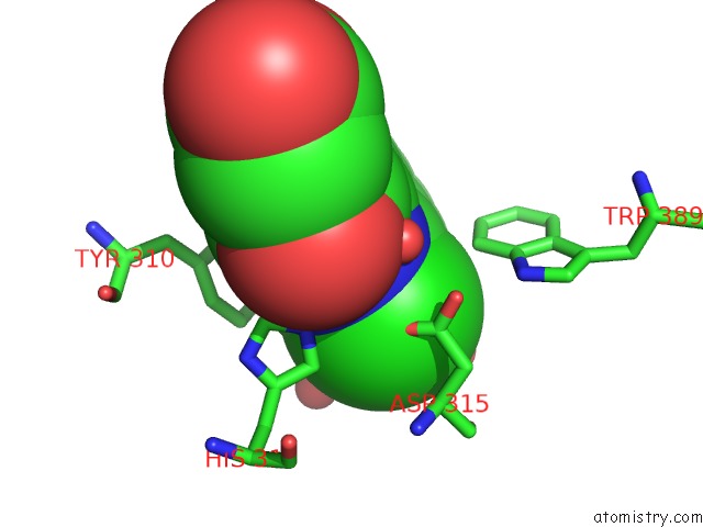
Mono view
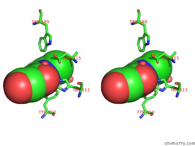
Stereo pair view
Mono view
Stereo pair view
|
|
A full contact list of Chlorine with other atoms in the Cl binding
site number 1 of Crystal Structure of Hif Prolyl Hydroxylase Egln-1 in Complex with A Biologically Active Inhibitor within 5.0Å range:
|
Reference:
A.G.Evdokimov,
R.L.Walter,
M.Mekel,
M.E.Pokross,
R.Kawamoto,
A.Boyer.
Crystal Structure of Hif Prolyl Hydroxylase in Complex with A Biologically Active Inhibitor To Be Published.
Page generated: Thu Jul 10 22:32:01 2025
Last articles
Zn in 8YFPZn in 8YB6
Zn in 8YF2
Zn in 8YAT
Zn in 8YAF
Zn in 8Y7E
Zn in 8Y9C
Zn in 8Y9B
Zn in 8Y6K
Zn in 8Y7W