Chlorine »
PDB 2hrc-2i6f »
2i58 »
Chlorine in PDB 2i58: Crystal Structure of Rafe From Streptococcus Pneumoniae Complexed with Raffinose
Protein crystallography data
The structure of Crystal Structure of Rafe From Streptococcus Pneumoniae Complexed with Raffinose, PDB code: 2i58
was solved by
N.G.Paterson,
A.Riboldi-Tunnicliffe,
T.J.Mitchell,
N.W.Isaacs,
with X-Ray Crystallography technique. A brief refinement statistics is given in the table below:
| Resolution Low / High (Å) | 46.27 / 2.80 |
| Space group | P 21 21 21 |
| Cell size a, b, c (Å), α, β, γ (°) | 49.214, 119.435, 146.326, 90.00, 90.00, 90.00 |
| R / Rfree (%) | 21.1 / 29.6 |
Chlorine Binding Sites:
The binding sites of Chlorine atom in the Crystal Structure of Rafe From Streptococcus Pneumoniae Complexed with Raffinose
(pdb code 2i58). This binding sites where shown within
5.0 Angstroms radius around Chlorine atom.
In total 3 binding sites of Chlorine where determined in the Crystal Structure of Rafe From Streptococcus Pneumoniae Complexed with Raffinose, PDB code: 2i58:
Jump to Chlorine binding site number: 1; 2; 3;
In total 3 binding sites of Chlorine where determined in the Crystal Structure of Rafe From Streptococcus Pneumoniae Complexed with Raffinose, PDB code: 2i58:
Jump to Chlorine binding site number: 1; 2; 3;
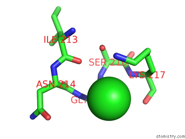
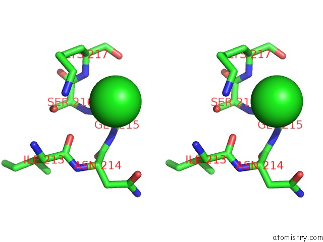
Chlorine binding site 1 out of 3 in 2i58
Go back to
Chlorine binding site 1 out
of 3 in the Crystal Structure of Rafe From Streptococcus Pneumoniae Complexed with Raffinose
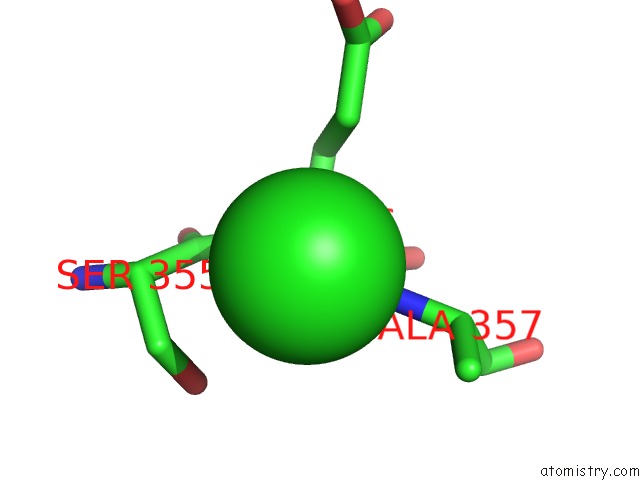
Mono view
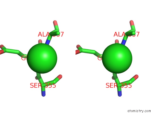
Stereo pair view
Mono view
Stereo pair view
|
|
A full contact list of Chlorine with other atoms in the Cl binding
site number 1 of Crystal Structure of Rafe From Streptococcus Pneumoniae Complexed with Raffinose within 5.0Å range:
|
Chlorine binding site 2 out of 3 in 2i58
Go back to
Chlorine binding site 2 out
of 3 in the Crystal Structure of Rafe From Streptococcus Pneumoniae Complexed with Raffinose
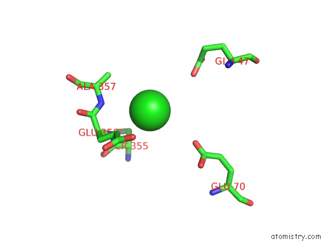
Mono view
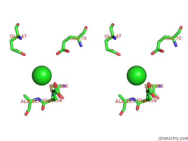
Stereo pair view
Mono view
Stereo pair view
|
|
A full contact list of Chlorine with other atoms in the Cl binding
site number 2 of Crystal Structure of Rafe From Streptococcus Pneumoniae Complexed with Raffinose within 5.0Å range:
|
Chlorine binding site 3 out of 3 in 2i58
Go back to
Chlorine binding site 3 out
of 3 in the Crystal Structure of Rafe From Streptococcus Pneumoniae Complexed with Raffinose
Mono view
Stereo pair view
Mono view
Stereo pair view
|
|
A full contact list of Chlorine with other atoms in the Cl binding
site number 3 of Crystal Structure of Rafe From Streptococcus Pneumoniae Complexed with Raffinose within 5.0Å range:
|
Reference:
N.G.Paterson,
A.Riboldi-Tunnicliffe,
T.J.Mitchell,
N.W.Isaacs.
Crystal Structure of Apo and Bound Forms of Rafe From Streptococcus Pneumoniae To Be Published.
Page generated: Thu Jul 10 22:41:11 2025
Last articles
Ni in 5Q5ENi in 5Q5C
Ni in 5Q5D
Ni in 5Q5B
Ni in 5Q5A
Ni in 5Q58
Ni in 5Q59
Ni in 5Q57
Ni in 5Q56
Ni in 5Q54