Chlorine »
PDB 2i6g-2ii3 »
2i9f »
Chlorine in PDB 2i9f: Structure of the Equine Arterivirus Nucleocapsid Protein
Protein crystallography data
The structure of Structure of the Equine Arterivirus Nucleocapsid Protein, PDB code: 2i9f
was solved by
A.Deshpande,
S.Wang,
M.Walsh,
T.Dokland,
with X-Ray Crystallography technique. A brief refinement statistics is given in the table below:
| Resolution Low / High (Å) | 49.94 / 2.00 |
| Space group | C 2 2 21 |
| Cell size a, b, c (Å), α, β, γ (°) | 68.230, 73.230, 138.960, 90.00, 90.00, 90.00 |
| R / Rfree (%) | 21.7 / 24.8 |
Other elements in 2i9f:
The structure of Structure of the Equine Arterivirus Nucleocapsid Protein also contains other interesting chemical elements:
| Sodium | (Na) | 2 atoms |
Chlorine Binding Sites:
The binding sites of Chlorine atom in the Structure of the Equine Arterivirus Nucleocapsid Protein
(pdb code 2i9f). This binding sites where shown within
5.0 Angstroms radius around Chlorine atom.
In total only one binding site of Chlorine was determined in the Structure of the Equine Arterivirus Nucleocapsid Protein, PDB code: 2i9f:
In total only one binding site of Chlorine was determined in the Structure of the Equine Arterivirus Nucleocapsid Protein, PDB code: 2i9f:
Chlorine binding site 1 out of 1 in 2i9f
Go back to
Chlorine binding site 1 out
of 1 in the Structure of the Equine Arterivirus Nucleocapsid Protein
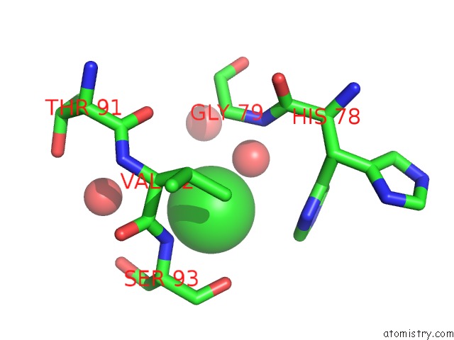
Mono view
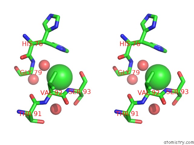
Stereo pair view
Mono view
Stereo pair view
A full contact list of Chlorine with other atoms in the Cl binding
site number 1 of Structure of the Equine Arterivirus Nucleocapsid Protein within 5.0Å range:
|
Reference:
A.Deshpande,
S.Wang,
M.A.Walsh,
T.Dokland.
Structure of the Equine Arteritis Virus Nucleocapsid Protein Reveals A Dimer-Dimer Arrangement. Acta Crystallogr.,Sect.D V. 63 581 2007.
ISSN: ISSN 0907-4449
PubMed: 17452783
DOI: 10.1107/S0907444907008372
Page generated: Thu Jul 10 22:42:21 2025
ISSN: ISSN 0907-4449
PubMed: 17452783
DOI: 10.1107/S0907444907008372
Last articles
Mg in 2A69Mg in 2A6E
Mg in 2A5Z
Mg in 2A5L
Mg in 2A5Y
Mg in 2A5J
Mg in 2A43
Mg in 2A5G
Mg in 2A5D
Mg in 2A5F