Chlorine »
PDB 2i6g-2ii3 »
2iay »
Chlorine in PDB 2iay: Crystal Structure of A DUF1831 Family Protein (LP2179) From Lactobacillus Plantarum at 1.20 A Resolution
Protein crystallography data
The structure of Crystal Structure of A DUF1831 Family Protein (LP2179) From Lactobacillus Plantarum at 1.20 A Resolution, PDB code: 2iay
was solved by
Joint Center For Structural Genomics (Jcsg),
with X-Ray Crystallography technique. A brief refinement statistics is given in the table below:
| Resolution Low / High (Å) | 28.93 / 1.20 |
| Space group | P 21 21 21 |
| Cell size a, b, c (Å), α, β, γ (°) | 36.289, 47.896, 58.010, 90.00, 90.00, 90.00 |
| R / Rfree (%) | 12 / 14.7 |
Chlorine Binding Sites:
The binding sites of Chlorine atom in the Crystal Structure of A DUF1831 Family Protein (LP2179) From Lactobacillus Plantarum at 1.20 A Resolution
(pdb code 2iay). This binding sites where shown within
5.0 Angstroms radius around Chlorine atom.
In total only one binding site of Chlorine was determined in the Crystal Structure of A DUF1831 Family Protein (LP2179) From Lactobacillus Plantarum at 1.20 A Resolution, PDB code: 2iay:
In total only one binding site of Chlorine was determined in the Crystal Structure of A DUF1831 Family Protein (LP2179) From Lactobacillus Plantarum at 1.20 A Resolution, PDB code: 2iay:
Chlorine binding site 1 out of 1 in 2iay
Go back to
Chlorine binding site 1 out
of 1 in the Crystal Structure of A DUF1831 Family Protein (LP2179) From Lactobacillus Plantarum at 1.20 A Resolution
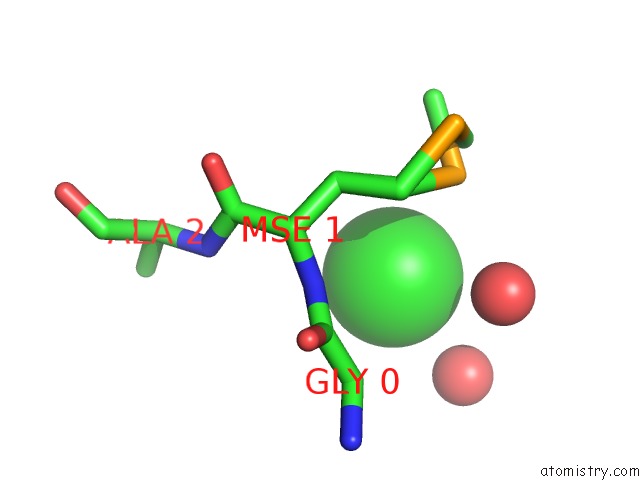
Mono view
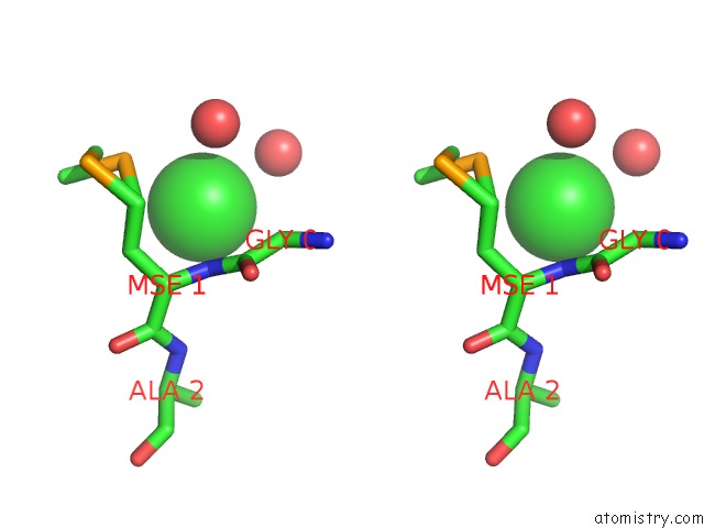
Stereo pair view
Mono view
Stereo pair view
A full contact list of Chlorine with other atoms in the Cl binding
site number 1 of Crystal Structure of A DUF1831 Family Protein (LP2179) From Lactobacillus Plantarum at 1.20 A Resolution within 5.0Å range:
|
Reference:
C.Bakolitsa,
A.Kumar,
D.Carlton,
M.D.Miller,
S.S.Krishna,
P.Abdubek,
T.Astakhova,
H.L.Axelrod,
H.J.Chiu,
T.Clayton,
M.C.Deller,
L.Duan,
M.A.Elsliger,
J.Feuerhelm,
S.K.Grzechnik,
J.C.Grant,
G.W.Han,
L.Jaroszewski,
K.K.Jin,
H.E.Klock,
M.W.Knuth,
P.Kozbial,
D.Marciano,
D.Mcmullan,
A.T.Morse,
E.Nigoghossian,
L.Okach,
S.Oommachen,
J.Paulsen,
R.Reyes,
C.L.Rife,
H.J.Tien,
C.V.Trout,
H.Van Den Bedem,
D.Weekes,
Q.Xu,
K.O.Hodgson,
J.Wooley,
A.M.Deacon,
A.Godzik,
S.A.Lesley,
I.A.Wilson.
Structure of LP2179, the First Representative of Pfam Family PF08866, Suggests A New Fold with A Role in Amino-Acid Metabolism. Acta Crystallogr.,Sect.F V. 66 1205 2010.
ISSN: ESSN 1744-3091
PubMed: 20944212
DOI: 10.1107/S1744309109023689
Page generated: Thu Jul 10 22:42:36 2025
ISSN: ESSN 1744-3091
PubMed: 20944212
DOI: 10.1107/S1744309109023689
Last articles
Mg in 5CDQMg in 5CGE
Mg in 5CGA
Mg in 5CG6
Mg in 5CG5
Mg in 5CFV
Mg in 5CFG
Mg in 5CFA
Mg in 5CC8
Mg in 5CEW