Chlorine »
PDB 2kug-2nwh »
2nn8 »
Chlorine in PDB 2nn8: Crystal Structure of Human Galectin-3 Carbohydrate-Recognition Domain with Lactose Bound, at 1.35 Angstrom Resolution
Protein crystallography data
The structure of Crystal Structure of Human Galectin-3 Carbohydrate-Recognition Domain with Lactose Bound, at 1.35 Angstrom Resolution, PDB code: 2nn8
was solved by
H.Blanchard,
P.M.Collins,
with X-Ray Crystallography technique. A brief refinement statistics is given in the table below:
| Resolution Low / High (Å) | 27.62 / 1.35 |
| Space group | P 21 21 21 |
| Cell size a, b, c (Å), α, β, γ (°) | 36.371, 57.800, 62.603, 90.00, 90.00, 90.00 |
| R / Rfree (%) | 16.4 / 17.5 |
Chlorine Binding Sites:
The binding sites of Chlorine atom in the Crystal Structure of Human Galectin-3 Carbohydrate-Recognition Domain with Lactose Bound, at 1.35 Angstrom Resolution
(pdb code 2nn8). This binding sites where shown within
5.0 Angstroms radius around Chlorine atom.
In total only one binding site of Chlorine was determined in the Crystal Structure of Human Galectin-3 Carbohydrate-Recognition Domain with Lactose Bound, at 1.35 Angstrom Resolution, PDB code: 2nn8:
In total only one binding site of Chlorine was determined in the Crystal Structure of Human Galectin-3 Carbohydrate-Recognition Domain with Lactose Bound, at 1.35 Angstrom Resolution, PDB code: 2nn8:
Chlorine binding site 1 out of 1 in 2nn8
Go back to
Chlorine binding site 1 out
of 1 in the Crystal Structure of Human Galectin-3 Carbohydrate-Recognition Domain with Lactose Bound, at 1.35 Angstrom Resolution

Mono view
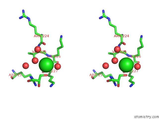
Stereo pair view
Mono view
Stereo pair view
A full contact list of Chlorine with other atoms in the Cl binding
site number 1 of Crystal Structure of Human Galectin-3 Carbohydrate-Recognition Domain with Lactose Bound, at 1.35 Angstrom Resolution within 5.0Å range:
|
Reference:
P.M.Collins,
K.I.Hidari,
H.Blanchard.
Slow Diffusion of Lactose Out of Galectin-3 Crystals Monitored By X-Ray Crystallography: Possible Implications For Ligand-Exchange Protocols. Acta Crystallogr.,Sect.D V. 63 415 2007.
ISSN: ISSN 0907-4449
PubMed: 17327679
DOI: 10.1107/S090744490605270X
Page generated: Thu Jul 10 23:18:35 2025
ISSN: ISSN 0907-4449
PubMed: 17327679
DOI: 10.1107/S090744490605270X
Last articles
Mg in 3T2CMg in 3T2B
Mg in 3T1R
Mg in 3T1O
Mg in 3T0D
Mg in 3T1Q
Mg in 3T12
Mg in 3T1K
Mg in 3T10
Mg in 3T0Z