Chlorine »
PDB 2nxw-2o9s »
2o0r »
Chlorine in PDB 2o0r: The Three-Dimensional Structure of N-Succinyldiaminopimelate Aminotransferase From Mycobacterium Tuberculosis
Protein crystallography data
The structure of The Three-Dimensional Structure of N-Succinyldiaminopimelate Aminotransferase From Mycobacterium Tuberculosis, PDB code: 2o0r
was solved by
S.Weyand,
G.Kefala,
M.S.Weiss,
Tb Structural Genomics Consortium (Tbsgc),
with X-Ray Crystallography technique. A brief refinement statistics is given in the table below:
| Resolution Low / High (Å) | 20.00 / 2.00 |
| Space group | P 2 2 21 |
| Cell size a, b, c (Å), α, β, γ (°) | 54.340, 56.130, 247.470, 90.00, 90.00, 90.00 |
| R / Rfree (%) | 16.4 / 21.3 |
Other elements in 2o0r:
The structure of The Three-Dimensional Structure of N-Succinyldiaminopimelate Aminotransferase From Mycobacterium Tuberculosis also contains other interesting chemical elements:
| Sodium | (Na) | 1 atom |
Chlorine Binding Sites:
The binding sites of Chlorine atom in the The Three-Dimensional Structure of N-Succinyldiaminopimelate Aminotransferase From Mycobacterium Tuberculosis
(pdb code 2o0r). This binding sites where shown within
5.0 Angstroms radius around Chlorine atom.
In total 2 binding sites of Chlorine where determined in the The Three-Dimensional Structure of N-Succinyldiaminopimelate Aminotransferase From Mycobacterium Tuberculosis, PDB code: 2o0r:
Jump to Chlorine binding site number: 1; 2;
In total 2 binding sites of Chlorine where determined in the The Three-Dimensional Structure of N-Succinyldiaminopimelate Aminotransferase From Mycobacterium Tuberculosis, PDB code: 2o0r:
Jump to Chlorine binding site number: 1; 2;
Chlorine binding site 1 out of 2 in 2o0r
Go back to
Chlorine binding site 1 out
of 2 in the The Three-Dimensional Structure of N-Succinyldiaminopimelate Aminotransferase From Mycobacterium Tuberculosis
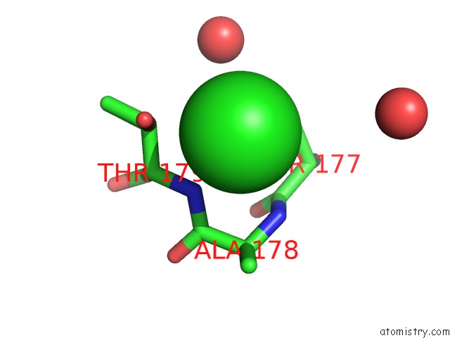
Mono view
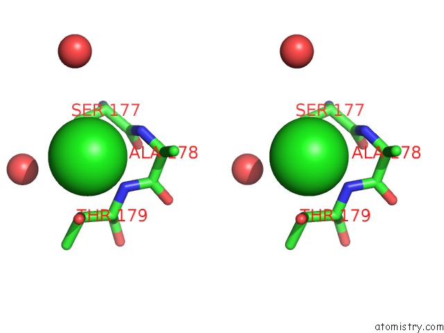
Stereo pair view
Mono view
Stereo pair view
A full contact list of Chlorine with other atoms in the Cl binding
site number 1 of The Three-Dimensional Structure of N-Succinyldiaminopimelate Aminotransferase From Mycobacterium Tuberculosis within 5.0Å range:
|
Chlorine binding site 2 out of 2 in 2o0r
Go back to
Chlorine binding site 2 out
of 2 in the The Three-Dimensional Structure of N-Succinyldiaminopimelate Aminotransferase From Mycobacterium Tuberculosis

Mono view
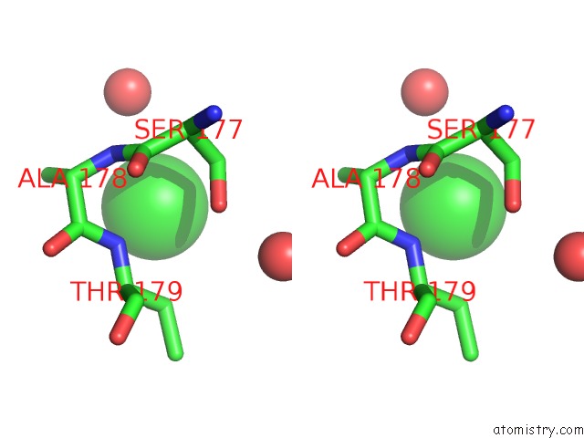
Stereo pair view
Mono view
Stereo pair view
A full contact list of Chlorine with other atoms in the Cl binding
site number 2 of The Three-Dimensional Structure of N-Succinyldiaminopimelate Aminotransferase From Mycobacterium Tuberculosis within 5.0Å range:
|
Reference:
S.Weyand,
G.Kefala,
M.S.Weiss.
The Three-Dimensional Structure of N-Succinyldiaminopimelate Aminotransferase From Mycobacterium Tuberculosis J.Mol.Biol. V. 367 825 2007.
ISSN: ISSN 0022-2836
PubMed: 17292400
DOI: 10.1016/J.JMB.2007.01.023
Page generated: Thu Jul 10 23:22:25 2025
ISSN: ISSN 0022-2836
PubMed: 17292400
DOI: 10.1016/J.JMB.2007.01.023
Last articles
Mg in 5WMBMg in 5WM8
Mg in 5WNO
Mg in 5WNI
Mg in 5WMT
Mg in 5WM1
Mg in 5WM6
Mg in 5WM4
Mg in 5WKC
Mg in 5WM3