Chlorine »
PDB 2ol4-2oud »
2om0 »
Chlorine in PDB 2om0: Structure of Human Insulin in Presence of Urea at pH 6.5
Protein crystallography data
The structure of Structure of Human Insulin in Presence of Urea at pH 6.5, PDB code: 2om0
was solved by
M.Norrman,
G.Schluckebier,
with X-Ray Crystallography technique. A brief refinement statistics is given in the table below:
| Resolution Low / High (Å) | 28.31 / 2.05 |
| Space group | C 2 2 21 1 |
| Cell size a, b, c (Å), α, β, γ (°) | 58.936, 219.318, 223.674, 90.00, 90.00, 90.00 |
| R / Rfree (%) | 18.4 / 22.7 |
Other elements in 2om0:
The structure of Structure of Human Insulin in Presence of Urea at pH 6.5 also contains other interesting chemical elements:
| Zinc | (Zn) | 6 atoms |
Chlorine Binding Sites:
The binding sites of Chlorine atom in the Structure of Human Insulin in Presence of Urea at pH 6.5
(pdb code 2om0). This binding sites where shown within
5.0 Angstroms radius around Chlorine atom.
In total 6 binding sites of Chlorine where determined in the Structure of Human Insulin in Presence of Urea at pH 6.5, PDB code: 2om0:
Jump to Chlorine binding site number: 1; 2; 3; 4; 5; 6;
In total 6 binding sites of Chlorine where determined in the Structure of Human Insulin in Presence of Urea at pH 6.5, PDB code: 2om0:
Jump to Chlorine binding site number: 1; 2; 3; 4; 5; 6;
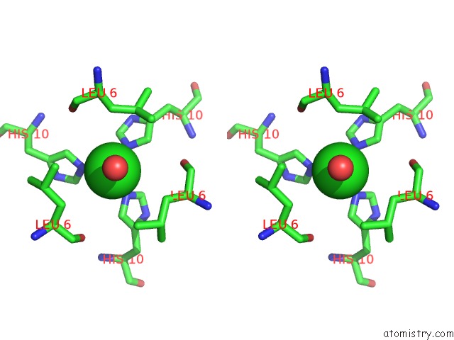
Chlorine binding site 1 out of 6 in 2om0
Go back to
Chlorine binding site 1 out
of 6 in the Structure of Human Insulin in Presence of Urea at pH 6.5
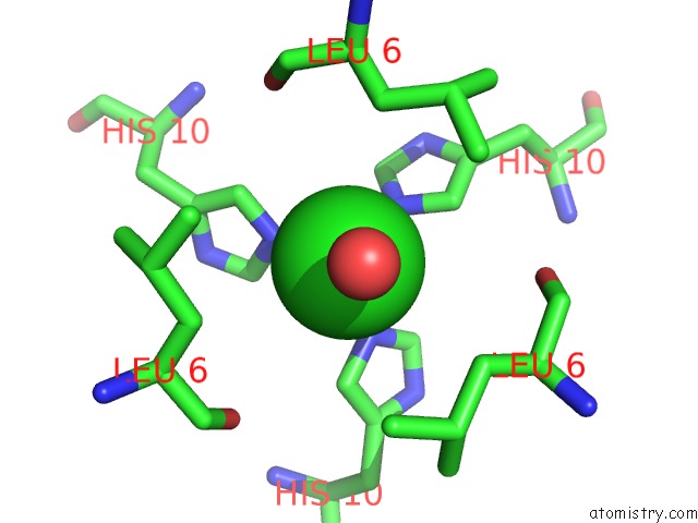
Mono view

Stereo pair view
Mono view
Stereo pair view
A full contact list of Chlorine with other atoms in the Cl binding
site number 1 of Structure of Human Insulin in Presence of Urea at pH 6.5 within 5.0Å range:
|
Chlorine binding site 2 out of 6 in 2om0
Go back to
Chlorine binding site 2 out
of 6 in the Structure of Human Insulin in Presence of Urea at pH 6.5
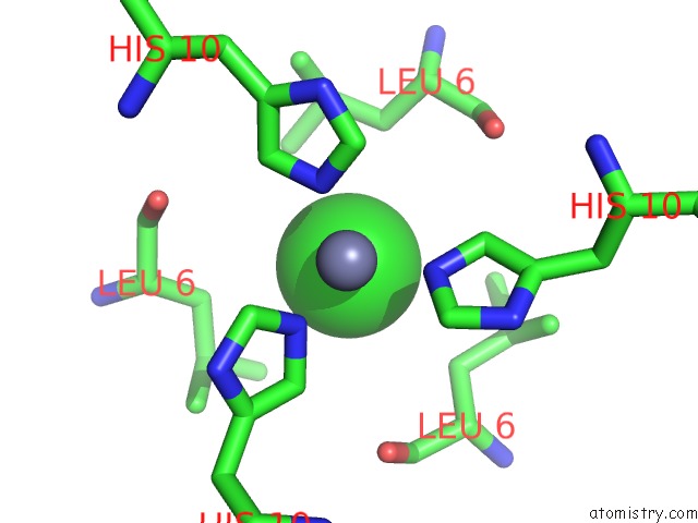
Mono view
Stereo pair view
Mono view
Stereo pair view
A full contact list of Chlorine with other atoms in the Cl binding
site number 2 of Structure of Human Insulin in Presence of Urea at pH 6.5 within 5.0Å range:
|
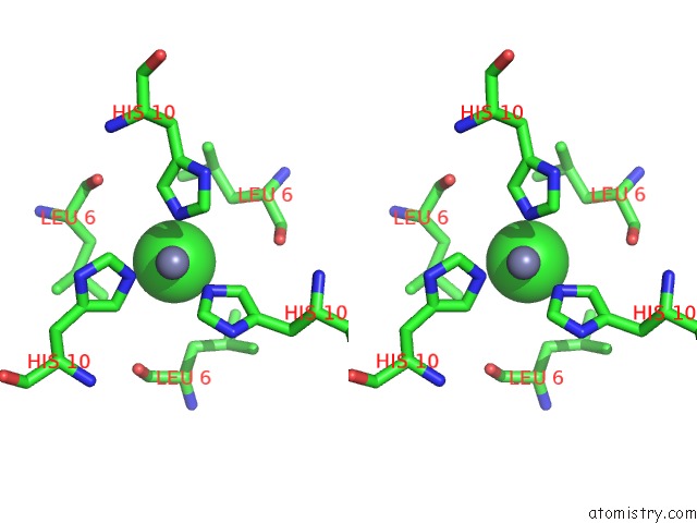
Chlorine binding site 3 out of 6 in 2om0
Go back to
Chlorine binding site 3 out
of 6 in the Structure of Human Insulin in Presence of Urea at pH 6.5
Mono view

Stereo pair view
Mono view
Stereo pair view
A full contact list of Chlorine with other atoms in the Cl binding
site number 3 of Structure of Human Insulin in Presence of Urea at pH 6.5 within 5.0Å range:
|
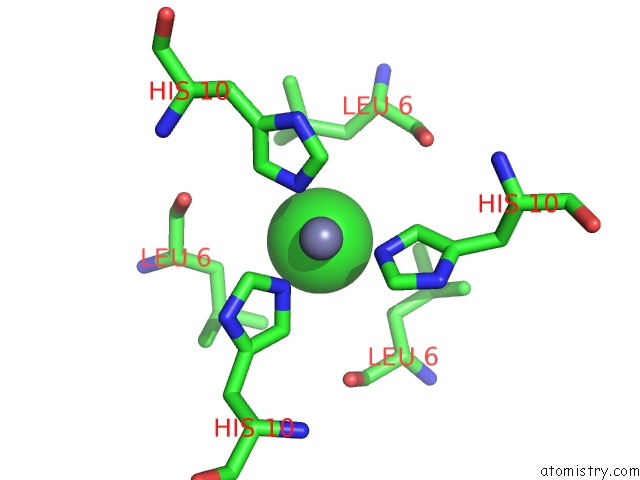
Chlorine binding site 4 out of 6 in 2om0
Go back to
Chlorine binding site 4 out
of 6 in the Structure of Human Insulin in Presence of Urea at pH 6.5

Mono view
Stereo pair view
Mono view
Stereo pair view
A full contact list of Chlorine with other atoms in the Cl binding
site number 4 of Structure of Human Insulin in Presence of Urea at pH 6.5 within 5.0Å range:
|
Chlorine binding site 5 out of 6 in 2om0
Go back to
Chlorine binding site 5 out
of 6 in the Structure of Human Insulin in Presence of Urea at pH 6.5

Mono view

Stereo pair view
Mono view
Stereo pair view
A full contact list of Chlorine with other atoms in the Cl binding
site number 5 of Structure of Human Insulin in Presence of Urea at pH 6.5 within 5.0Å range:
|
Chlorine binding site 6 out of 6 in 2om0
Go back to
Chlorine binding site 6 out
of 6 in the Structure of Human Insulin in Presence of Urea at pH 6.5

Mono view

Stereo pair view
Mono view
Stereo pair view
A full contact list of Chlorine with other atoms in the Cl binding
site number 6 of Structure of Human Insulin in Presence of Urea at pH 6.5 within 5.0Å range:
|
Reference:
M.Norrman,
G.Schluckebier.
Crystallographic Characterization of Two Novel Crystal Forms of Human Insulin Induced By Chaotropic Agents and A Shift in pH. Bmc Struct.Biol. V. 7 83 2007.
ISSN: ESSN 1472-6807
PubMed: 18093308
DOI: 10.1186/1472-6807-7-83
Page generated: Thu Jul 10 23:33:12 2025
ISSN: ESSN 1472-6807
PubMed: 18093308
DOI: 10.1186/1472-6807-7-83
Last articles
Mg in 8YEBMg in 8YFR
Mg in 8YFQ
Mg in 8YFP
Mg in 8YFN
Mg in 8YFK
Mg in 8YD1
Mg in 8YCX
Mg in 8YE0
Mg in 8YD0