Chlorine »
PDB 2ol4-2oud »
2oqx »
Chlorine in PDB 2oqx: Crystal Structure of the Apo Form of E. Coli Tryptophanase at 1.9 A Resolution
Enzymatic activity of Crystal Structure of the Apo Form of E. Coli Tryptophanase at 1.9 A Resolution
All present enzymatic activity of Crystal Structure of the Apo Form of E. Coli Tryptophanase at 1.9 A Resolution:
4.1.99.1;
4.1.99.1;
Protein crystallography data
The structure of Crystal Structure of the Apo Form of E. Coli Tryptophanase at 1.9 A Resolution, PDB code: 2oqx
was solved by
Y.Goldgur,
A.Kogan,
G.Gdalevsky,
A.Parola,
R.Cohen-Luria,
O.Almog,
with X-Ray Crystallography technique. A brief refinement statistics is given in the table below:
| Resolution Low / High (Å) | 14.82 / 1.90 |
| Space group | F 2 2 2 |
| Cell size a, b, c (Å), α, β, γ (°) | 118.422, 120.116, 171.209, 90.00, 90.00, 90.00 |
| R / Rfree (%) | 20.3 / 23.2 |
Other elements in 2oqx:
The structure of Crystal Structure of the Apo Form of E. Coli Tryptophanase at 1.9 A Resolution also contains other interesting chemical elements:
| Magnesium | (Mg) | 1 atom |
Chlorine Binding Sites:
The binding sites of Chlorine atom in the Crystal Structure of the Apo Form of E. Coli Tryptophanase at 1.9 A Resolution
(pdb code 2oqx). This binding sites where shown within
5.0 Angstroms radius around Chlorine atom.
In total only one binding site of Chlorine was determined in the Crystal Structure of the Apo Form of E. Coli Tryptophanase at 1.9 A Resolution, PDB code: 2oqx:
In total only one binding site of Chlorine was determined in the Crystal Structure of the Apo Form of E. Coli Tryptophanase at 1.9 A Resolution, PDB code: 2oqx:
Chlorine binding site 1 out of 1 in 2oqx
Go back to
Chlorine binding site 1 out
of 1 in the Crystal Structure of the Apo Form of E. Coli Tryptophanase at 1.9 A Resolution
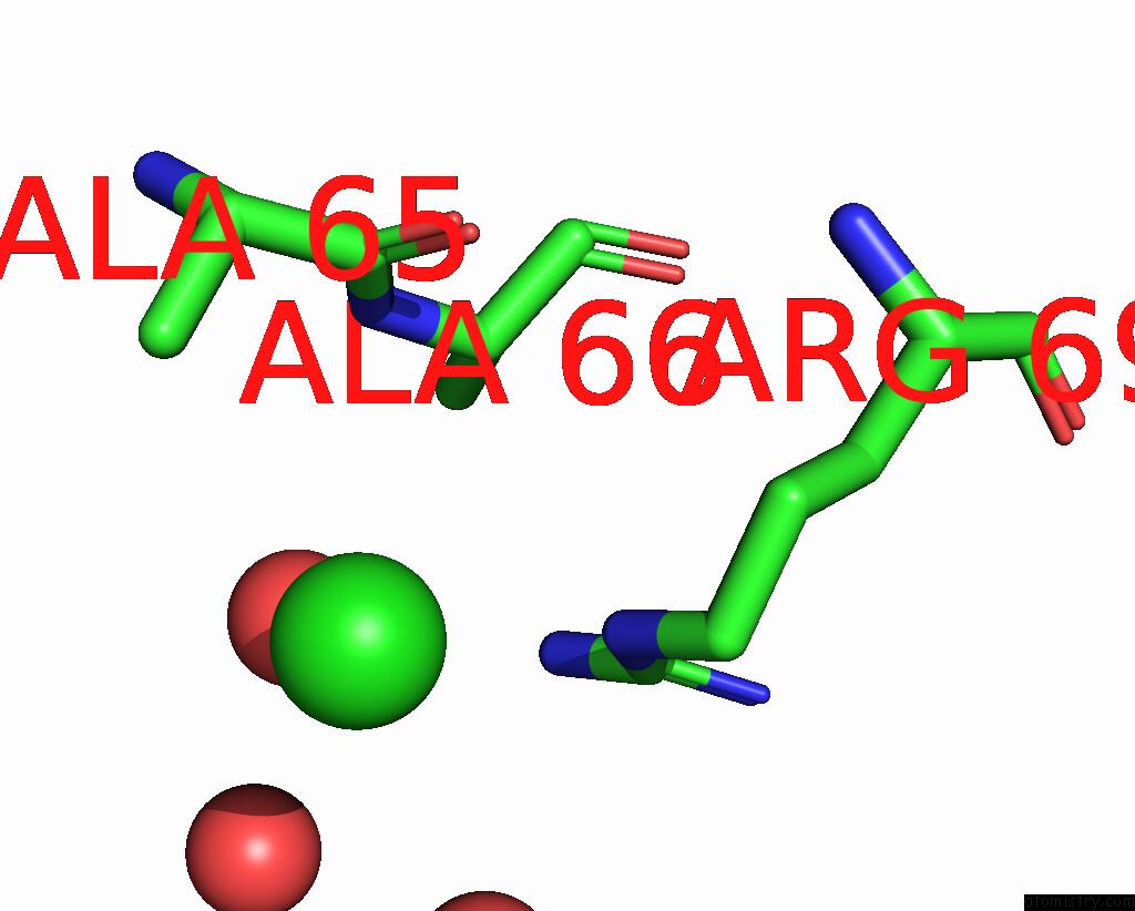
Mono view
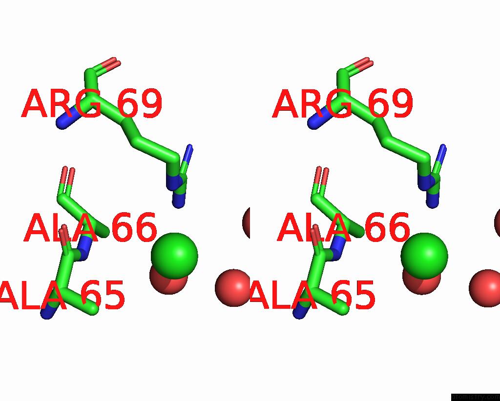
Stereo pair view
Mono view
Stereo pair view
A full contact list of Chlorine with other atoms in the Cl binding
site number 1 of Crystal Structure of the Apo Form of E. Coli Tryptophanase at 1.9 A Resolution within 5.0Å range:
|
Reference:
N.Tsesin,
A.Kogan,
G.Y.Gdalevsky,
J.P.Himanen,
R.Cohen-Luria,
A.H.Parola,
Y.Goldgur,
O.Almog.
The Structure of Apo Tryptophanase From Escherichia Coli Reveals A Wide-Open Conformation. Acta Crystallogr.,Sect.D V. 63 969 2007.
ISSN: ISSN 0907-4449
PubMed: 17704565
DOI: 10.1107/S0907444907036396
Page generated: Thu Jul 10 23:35:55 2025
ISSN: ISSN 0907-4449
PubMed: 17704565
DOI: 10.1107/S0907444907036396
Last articles
Mg in 6MXDMg in 6MXB
Mg in 6MW7
Mg in 6MWK
Mg in 6MTI
Mg in 6MVE
Mg in 6MV9
Mg in 6MU4
Mg in 6MTH
Mg in 6MTA