Chlorine »
PDB 2pgc-2px2 »
2pk0 »
Chlorine in PDB 2pk0: Structure of the S. Agalactiae Serine/Threonine Phosphatase at 2.65 Resolution
Enzymatic activity of Structure of the S. Agalactiae Serine/Threonine Phosphatase at 2.65 Resolution
All present enzymatic activity of Structure of the S. Agalactiae Serine/Threonine Phosphatase at 2.65 Resolution:
3.1.3.16;
3.1.3.16;
Protein crystallography data
The structure of Structure of the S. Agalactiae Serine/Threonine Phosphatase at 2.65 Resolution, PDB code: 2pk0
was solved by
M.K.Rantanen,
L.Lehtio,
L.Rajagopal,
C.E.Rubens,
A.Goldman,
with X-Ray Crystallography technique. A brief refinement statistics is given in the table below:
| Resolution Low / High (Å) | 19.65 / 2.65 |
| Space group | P 21 21 2 |
| Cell size a, b, c (Å), α, β, γ (°) | 139.400, 92.100, 86.900, 90.00, 90.00, 90.00 |
| R / Rfree (%) | 19.7 / 27.1 |
Other elements in 2pk0:
The structure of Structure of the S. Agalactiae Serine/Threonine Phosphatase at 2.65 Resolution also contains other interesting chemical elements:
| Magnesium | (Mg) | 10 atoms |
Chlorine Binding Sites:
The binding sites of Chlorine atom in the Structure of the S. Agalactiae Serine/Threonine Phosphatase at 2.65 Resolution
(pdb code 2pk0). This binding sites where shown within
5.0 Angstroms radius around Chlorine atom.
In total only one binding site of Chlorine was determined in the Structure of the S. Agalactiae Serine/Threonine Phosphatase at 2.65 Resolution, PDB code: 2pk0:
In total only one binding site of Chlorine was determined in the Structure of the S. Agalactiae Serine/Threonine Phosphatase at 2.65 Resolution, PDB code: 2pk0:
Chlorine binding site 1 out of 1 in 2pk0
Go back to
Chlorine binding site 1 out
of 1 in the Structure of the S. Agalactiae Serine/Threonine Phosphatase at 2.65 Resolution
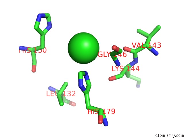
Mono view
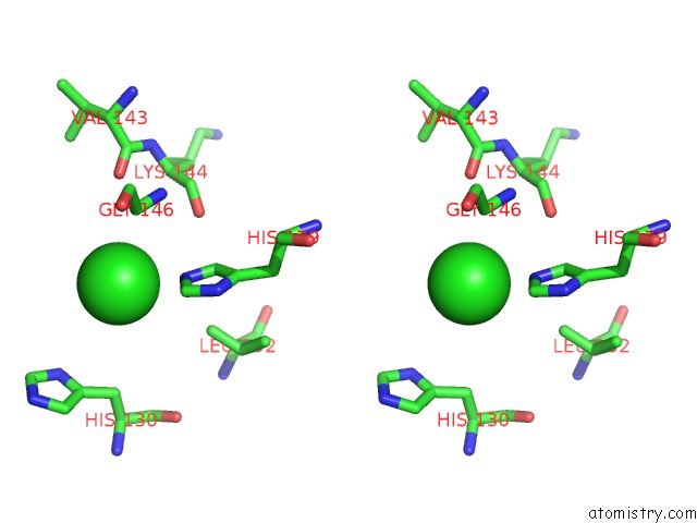
Stereo pair view
Mono view
Stereo pair view
|
|
A full contact list of Chlorine with other atoms in the Cl binding
site number 1 of Structure of the S. Agalactiae Serine/Threonine Phosphatase at 2.65 Resolution within 5.0Å range:
|
Reference:
M.K.Rantanen,
L.Lehtio,
L.Rajagopal,
C.E.Rubens,
A.Goldman.
Structure of Streptococcus Agalactiae Serine/Threonine Phosphatase. the Subdomain Conformation Is Coupled to the Binding of A Third Metal Ion Febs J. V. 274 3128 2007.
ISSN: ISSN 1742-464X
PubMed: 17521332
DOI: 10.1111/J.1742-4658.2007.05845.X
Page generated: Thu Jul 10 23:52:24 2025
ISSN: ISSN 1742-464X
PubMed: 17521332
DOI: 10.1111/J.1742-4658.2007.05845.X
Last articles
Fe in 2YXOFe in 2YRS
Fe in 2YXC
Fe in 2YNM
Fe in 2YVJ
Fe in 2YP1
Fe in 2YU2
Fe in 2YU1
Fe in 2YQB
Fe in 2YOO