Chlorine »
PDB 2px4-2q6r »
2px8 »
Chlorine in PDB 2px8: Crystal Structure of the Murray Valley Encephalitis Virus NS5 2'-O Methyltransferase Domain in Complex with Sah and 7M-Gtp
Enzymatic activity of Crystal Structure of the Murray Valley Encephalitis Virus NS5 2'-O Methyltransferase Domain in Complex with Sah and 7M-Gtp
All present enzymatic activity of Crystal Structure of the Murray Valley Encephalitis Virus NS5 2'-O Methyltransferase Domain in Complex with Sah and 7M-Gtp:
2.7.7.48;
2.7.7.48;
Protein crystallography data
The structure of Crystal Structure of the Murray Valley Encephalitis Virus NS5 2'-O Methyltransferase Domain in Complex with Sah and 7M-Gtp, PDB code: 2px8
was solved by
R.Assenberg,
J.Ren,
A.Verma,
T.S.Walter,
D.Alderton,
R.J.Hurrelbrink,
S.D.Fuller,
R.J.Owens,
D.I.Stuart,
J.M.Grimes,
Oxford Protein Productionfacility (Oppf),
with X-Ray Crystallography technique. A brief refinement statistics is given in the table below:
| Resolution Low / High (Å) | 30.00 / 2.20 |
| Space group | P 21 21 21 |
| Cell size a, b, c (Å), α, β, γ (°) | 67.579, 87.445, 100.914, 90.00, 90.00, 90.00 |
| R / Rfree (%) | 16.6 / 21.7 |
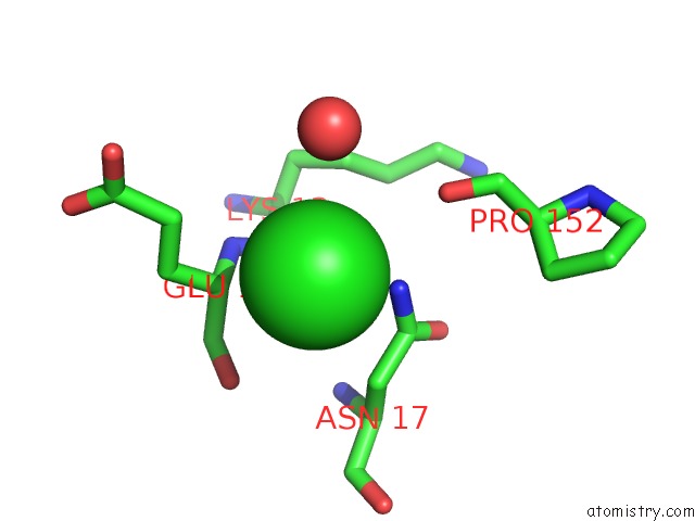
Chlorine Binding Sites:
The binding sites of Chlorine atom in the Crystal Structure of the Murray Valley Encephalitis Virus NS5 2'-O Methyltransferase Domain in Complex with Sah and 7M-Gtp
(pdb code 2px8). This binding sites where shown within
5.0 Angstroms radius around Chlorine atom.
In total only one binding site of Chlorine was determined in the Crystal Structure of the Murray Valley Encephalitis Virus NS5 2'-O Methyltransferase Domain in Complex with Sah and 7M-Gtp, PDB code: 2px8:
In total only one binding site of Chlorine was determined in the Crystal Structure of the Murray Valley Encephalitis Virus NS5 2'-O Methyltransferase Domain in Complex with Sah and 7M-Gtp, PDB code: 2px8:
Chlorine binding site 1 out of 1 in 2px8
Go back to
Chlorine binding site 1 out
of 1 in the Crystal Structure of the Murray Valley Encephalitis Virus NS5 2'-O Methyltransferase Domain in Complex with Sah and 7M-Gtp
Mono view
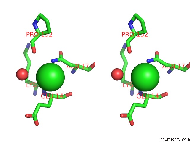
Stereo pair view
Mono view
Stereo pair view
A full contact list of Chlorine with other atoms in the Cl binding
site number 1 of Crystal Structure of the Murray Valley Encephalitis Virus NS5 2'-O Methyltransferase Domain in Complex with Sah and 7M-Gtp within 5.0Å range:
|
Reference:
R.Assenberg,
J.Ren,
A.Verma,
T.S.Walter,
D.Alderton,
R.J.Hurrelbrink,
S.D.Fuller,
S.Bressanelli,
R.J.Owens,
D.I.Stuart,
J.M.Grimes.
Crystal Structure of the Murray Valley Encephalitis Virus NS5 Methyltransferase Domain in Complex with Cap Analogues. J.Gen.Virol. V. 88 2228 2007.
ISSN: ISSN 0022-1317
PubMed: 17622627
DOI: 10.1099/VIR.0.82757-0
Page generated: Thu Jul 10 23:57:46 2025
ISSN: ISSN 0022-1317
PubMed: 17622627
DOI: 10.1099/VIR.0.82757-0
Last articles
Fe in 8Z9SFe in 8Z1Z
Fe in 8Z4Q
Fe in 8YS5
Fe in 8Z1Y
Fe in 8Z1X
Fe in 8Z11
Fe in 8Z1W
Fe in 8Z1V
Fe in 8YZ8