Chlorine »
PDB 2qio-2qu5 »
2qsq »
Chlorine in PDB 2qsq: Crystal Structure of the N-Terminal Domain of Carcinoembryonic Antigen (Cea)
Protein crystallography data
The structure of Crystal Structure of the N-Terminal Domain of Carcinoembryonic Antigen (Cea), PDB code: 2qsq
was solved by
I.Le Trong,
N.Korotkova,
S.L.Moseley,
R.E.Stenkamp,
with X-Ray Crystallography technique. A brief refinement statistics is given in the table below:
| Resolution Low / High (Å) | 10.00 / 1.95 |
| Space group | H 3 |
| Cell size a, b, c (Å), α, β, γ (°) | 132.531, 132.531, 82.762, 90.00, 90.00, 120.00 |
| R / Rfree (%) | 18 / 20.3 |
Chlorine Binding Sites:
The binding sites of Chlorine atom in the Crystal Structure of the N-Terminal Domain of Carcinoembryonic Antigen (Cea)
(pdb code 2qsq). This binding sites where shown within
5.0 Angstroms radius around Chlorine atom.
In total 2 binding sites of Chlorine where determined in the Crystal Structure of the N-Terminal Domain of Carcinoembryonic Antigen (Cea), PDB code: 2qsq:
Jump to Chlorine binding site number: 1; 2;
In total 2 binding sites of Chlorine where determined in the Crystal Structure of the N-Terminal Domain of Carcinoembryonic Antigen (Cea), PDB code: 2qsq:
Jump to Chlorine binding site number: 1; 2;
Chlorine binding site 1 out of 2 in 2qsq
Go back to
Chlorine binding site 1 out
of 2 in the Crystal Structure of the N-Terminal Domain of Carcinoembryonic Antigen (Cea)
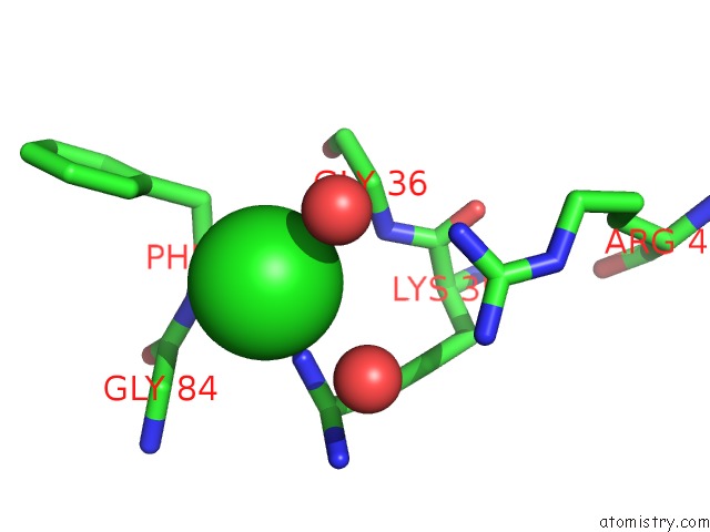
Mono view
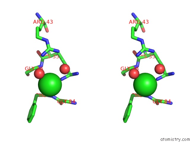
Stereo pair view
Mono view
Stereo pair view
A full contact list of Chlorine with other atoms in the Cl binding
site number 1 of Crystal Structure of the N-Terminal Domain of Carcinoembryonic Antigen (Cea) within 5.0Å range:
|
Chlorine binding site 2 out of 2 in 2qsq
Go back to
Chlorine binding site 2 out
of 2 in the Crystal Structure of the N-Terminal Domain of Carcinoembryonic Antigen (Cea)
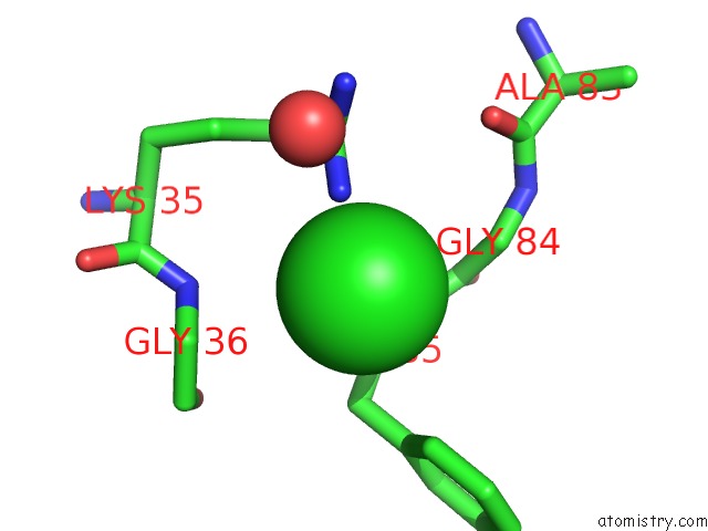
Mono view
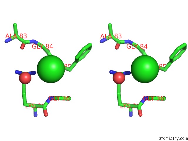
Stereo pair view
Mono view
Stereo pair view
A full contact list of Chlorine with other atoms in the Cl binding
site number 2 of Crystal Structure of the N-Terminal Domain of Carcinoembryonic Antigen (Cea) within 5.0Å range:
|
Reference:
N.Korotkova,
Y.Yang,
I.Le Trong,
E.Cota,
B.Demeler,
J.Marchant,
W.E.Thomas,
R.E.Stenkamp,
S.L.Moseley,
S.Matthews.
Binding of Dr Adhesins of Escherichia Coli to Carcinoembryonic Antigen Triggers Receptor Dissociation. Mol.Microbiol. V. 67 420 2008.
ISSN: ISSN 0950-382X
PubMed: 18086185
DOI: 10.1111/J.1365-2958.2007.06054.X
Page generated: Fri Jul 11 00:17:27 2025
ISSN: ISSN 0950-382X
PubMed: 18086185
DOI: 10.1111/J.1365-2958.2007.06054.X
Last articles
Mg in 4G6HMg in 4G61
Mg in 4G4S
Mg in 4G5Y
Mg in 4G5G
Mg in 4G3S
Mg in 4G3D
Mg in 4G3X
Mg in 4G3P
Mg in 4G3Q