Chlorine »
PDB 2r7k-2rh8 »
2rfv »
Chlorine in PDB 2rfv: High Resolution Structure of L-Methionine Gamma-Lyase From Citrobacter Freundii
Enzymatic activity of High Resolution Structure of L-Methionine Gamma-Lyase From Citrobacter Freundii
All present enzymatic activity of High Resolution Structure of L-Methionine Gamma-Lyase From Citrobacter Freundii:
4.4.1.11;
4.4.1.11;
Protein crystallography data
The structure of High Resolution Structure of L-Methionine Gamma-Lyase From Citrobacter Freundii, PDB code: 2rfv
was solved by
A.D.Nikulin,
S.V.Revtovich,
E.A.Morozova,
N.A.Nevskaya,
S.V.Nikonov,
M.B.Garber,
T.V.Demidkina,
with X-Ray Crystallography technique. A brief refinement statistics is given in the table below:
| Resolution Low / High (Å) | 20.00 / 1.36 |
| Space group | I 2 2 2 |
| Cell size a, b, c (Å), α, β, γ (°) | 56.450, 122.800, 127.920, 90.00, 90.00, 90.00 |
| R / Rfree (%) | 15.2 / 17.7 |
Chlorine Binding Sites:
The binding sites of Chlorine atom in the High Resolution Structure of L-Methionine Gamma-Lyase From Citrobacter Freundii
(pdb code 2rfv). This binding sites where shown within
5.0 Angstroms radius around Chlorine atom.
In total only one binding site of Chlorine was determined in the High Resolution Structure of L-Methionine Gamma-Lyase From Citrobacter Freundii, PDB code: 2rfv:
In total only one binding site of Chlorine was determined in the High Resolution Structure of L-Methionine Gamma-Lyase From Citrobacter Freundii, PDB code: 2rfv:
Chlorine binding site 1 out of 1 in 2rfv
Go back to
Chlorine binding site 1 out
of 1 in the High Resolution Structure of L-Methionine Gamma-Lyase From Citrobacter Freundii
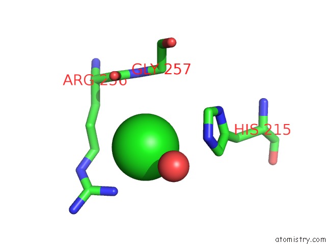
Mono view
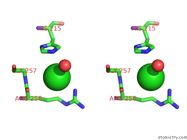
Stereo pair view
Mono view
Stereo pair view
A full contact list of Chlorine with other atoms in the Cl binding
site number 1 of High Resolution Structure of L-Methionine Gamma-Lyase From Citrobacter Freundii within 5.0Å range:
|
Reference:
A.Nikulin,
S.Revtovich,
E.Morozova,
N.Nevskaya,
S.Nikonov,
M.Garber,
T.Demidkina.
High-Resolution Structure of Methionine Gamma-Lyase From Citrobacter Freundii. Acta Crystallogr.,Sect.D V. 64 211 2008.
ISSN: ISSN 0907-4449
PubMed: 18219122
DOI: 10.1107/S0907444907065390
Page generated: Fri Jul 11 00:30:06 2025
ISSN: ISSN 0907-4449
PubMed: 18219122
DOI: 10.1107/S0907444907065390
Last articles
Mg in 6JNXMg in 6JMG
Mg in 6JNL
Mg in 6JLV
Mg in 6JLN
Mg in 6JLL
Mg in 6JLK
Mg in 6JLM
Mg in 6JLJ
Mg in 6JIL