Chlorine »
PDB 2rha-2uxn »
2rir »
Chlorine in PDB 2rir: Crystal Structure of Dipicolinate Synthase, A Chain, From Bacillus Subtilis
Protein crystallography data
The structure of Crystal Structure of Dipicolinate Synthase, A Chain, From Bacillus Subtilis, PDB code: 2rir
was solved by
J.Osipiuk,
P.Quartey,
S.Moy,
A.Joachimiak,
Midwest Center For Structuralgenomics (Mcsg),
with X-Ray Crystallography technique. A brief refinement statistics is given in the table below:
| Resolution Low / High (Å) | 40.00 / 2.79 |
| Space group | C 1 2 1 |
| Cell size a, b, c (Å), α, β, γ (°) | 261.578, 75.108, 200.464, 90.00, 119.74, 90.00 |
| R / Rfree (%) | 19.9 / 24.9 |
Chlorine Binding Sites:
The binding sites of Chlorine atom in the Crystal Structure of Dipicolinate Synthase, A Chain, From Bacillus Subtilis
(pdb code 2rir). This binding sites where shown within
5.0 Angstroms radius around Chlorine atom.
In total 7 binding sites of Chlorine where determined in the Crystal Structure of Dipicolinate Synthase, A Chain, From Bacillus Subtilis, PDB code: 2rir:
Jump to Chlorine binding site number: 1; 2; 3; 4; 5; 6; 7;
In total 7 binding sites of Chlorine where determined in the Crystal Structure of Dipicolinate Synthase, A Chain, From Bacillus Subtilis, PDB code: 2rir:
Jump to Chlorine binding site number: 1; 2; 3; 4; 5; 6; 7;
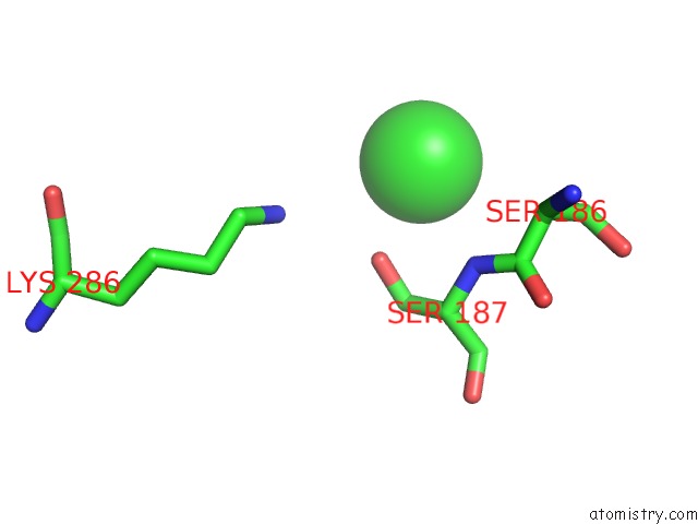
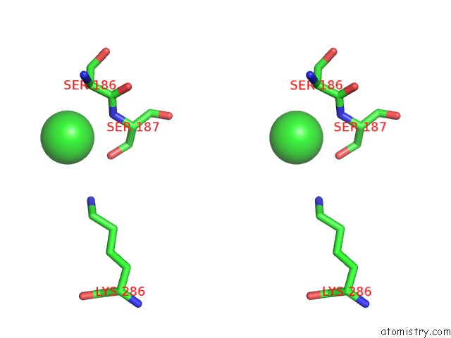
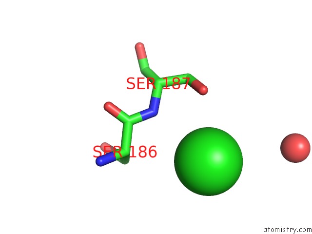
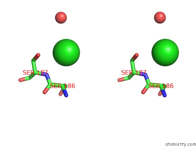
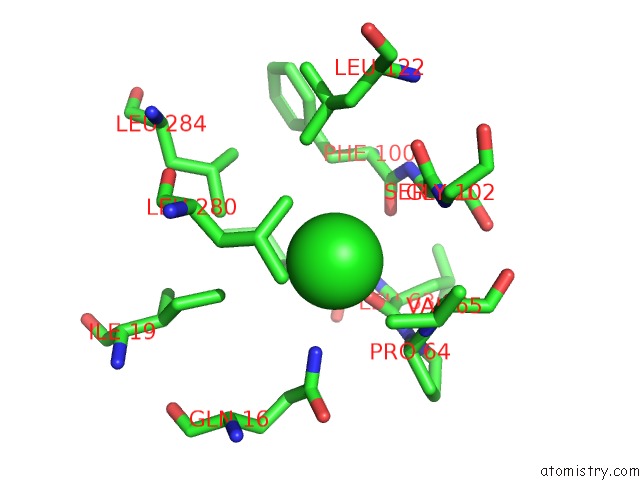
Chlorine binding site 1 out of 7 in 2rir
Go back to
Chlorine binding site 1 out
of 7 in the Crystal Structure of Dipicolinate Synthase, A Chain, From Bacillus Subtilis
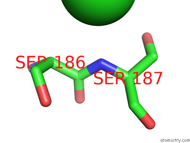
Mono view
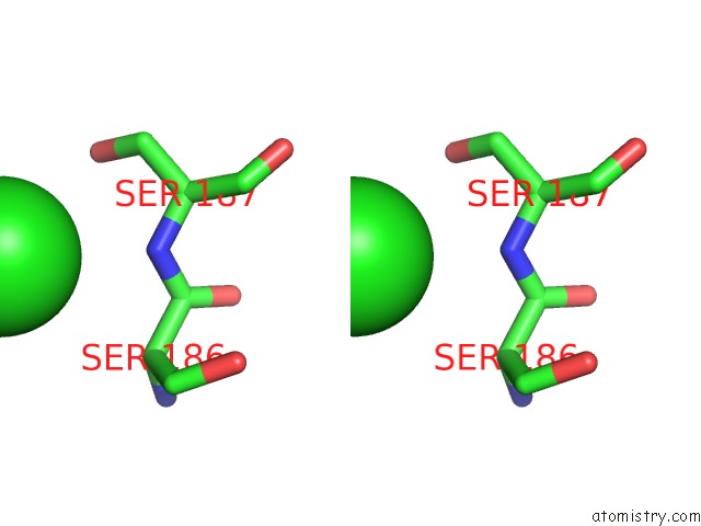
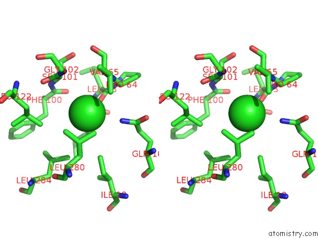
Stereo pair view
Mono view
Stereo pair view
A full contact list of Chlorine with other atoms in the Cl binding
site number 1 of Crystal Structure of Dipicolinate Synthase, A Chain, From Bacillus Subtilis within 5.0Å range:
|
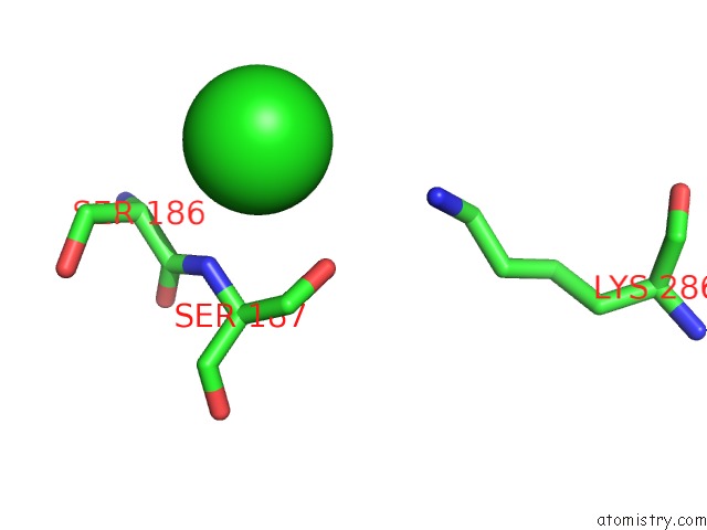
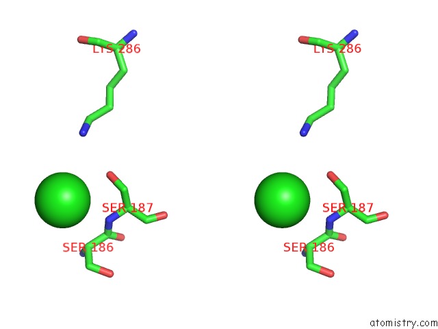
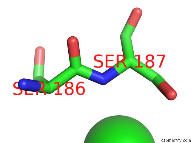
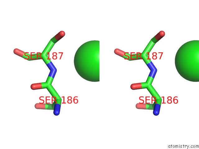
Chlorine binding site 2 out of 7 in 2rir
Go back to
Chlorine binding site 2 out
of 7 in the Crystal Structure of Dipicolinate Synthase, A Chain, From Bacillus Subtilis
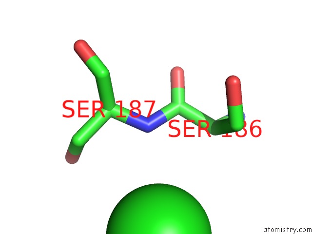
Mono view
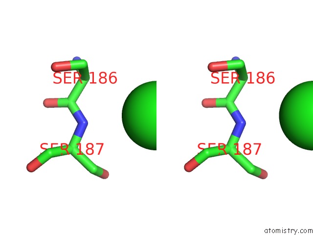
Stereo pair view
Mono view
Stereo pair view
A full contact list of Chlorine with other atoms in the Cl binding
site number 2 of Crystal Structure of Dipicolinate Synthase, A Chain, From Bacillus Subtilis within 5.0Å range:
|
Chlorine binding site 3 out of 7 in 2rir
Go back to
Chlorine binding site 3 out
of 7 in the Crystal Structure of Dipicolinate Synthase, A Chain, From Bacillus Subtilis
Mono view
Stereo pair view
Mono view
Stereo pair view
A full contact list of Chlorine with other atoms in the Cl binding
site number 3 of Crystal Structure of Dipicolinate Synthase, A Chain, From Bacillus Subtilis within 5.0Å range:
|
Chlorine binding site 4 out of 7 in 2rir
Go back to
Chlorine binding site 4 out
of 7 in the Crystal Structure of Dipicolinate Synthase, A Chain, From Bacillus Subtilis
Mono view
Stereo pair view
Mono view
Stereo pair view
A full contact list of Chlorine with other atoms in the Cl binding
site number 4 of Crystal Structure of Dipicolinate Synthase, A Chain, From Bacillus Subtilis within 5.0Å range:
|
Chlorine binding site 5 out of 7 in 2rir
Go back to
Chlorine binding site 5 out
of 7 in the Crystal Structure of Dipicolinate Synthase, A Chain, From Bacillus Subtilis
Mono view
Stereo pair view
Mono view
Stereo pair view
A full contact list of Chlorine with other atoms in the Cl binding
site number 5 of Crystal Structure of Dipicolinate Synthase, A Chain, From Bacillus Subtilis within 5.0Å range:
|
Chlorine binding site 6 out of 7 in 2rir
Go back to
Chlorine binding site 6 out
of 7 in the Crystal Structure of Dipicolinate Synthase, A Chain, From Bacillus Subtilis
Mono view
Stereo pair view
Mono view
Stereo pair view
A full contact list of Chlorine with other atoms in the Cl binding
site number 6 of Crystal Structure of Dipicolinate Synthase, A Chain, From Bacillus Subtilis within 5.0Å range:
|
Chlorine binding site 7 out of 7 in 2rir
Go back to
Chlorine binding site 7 out
of 7 in the Crystal Structure of Dipicolinate Synthase, A Chain, From Bacillus Subtilis
Mono view
Stereo pair view
Mono view
Stereo pair view
A full contact list of Chlorine with other atoms in the Cl binding
site number 7 of Crystal Structure of Dipicolinate Synthase, A Chain, From Bacillus Subtilis within 5.0Å range:
|
Reference:
J.Osipiuk,
P.Quartey,
S.Moy,
A.Joachimiak.
Crystal Structure of Dipicolinate Synthase, A Chain, From Bacillus Subtilis. To Be Published.
Page generated: Fri Jul 11 00:30:59 2025
Last articles
Mg in 6ZNDMg in 6ZNW
Mg in 6ZJB
Mg in 6ZN7
Mg in 6ZN4
Mg in 6ZMD
Mg in 6ZLI
Mg in 6ZM2
Mg in 6ZL7
Mg in 6ZL5