Chlorine »
PDB 2rha-2uxn »
2rj2 »
Chlorine in PDB 2rj2: Crystal Structure of the Sugar Recognizing Scf Ubiquitin Ligase at 1.7 Resolution
Enzymatic activity of Crystal Structure of the Sugar Recognizing Scf Ubiquitin Ligase at 1.7 Resolution
All present enzymatic activity of Crystal Structure of the Sugar Recognizing Scf Ubiquitin Ligase at 1.7 Resolution:
6.3.2.19;
6.3.2.19;
Protein crystallography data
The structure of Crystal Structure of the Sugar Recognizing Scf Ubiquitin Ligase at 1.7 Resolution, PDB code: 2rj2
was solved by
S.Vaijayanthimala,
D.Velmurugan,
T.Mizushima,
T.Yamane,
Y.Yoshida,
K.Tanaka,
with X-Ray Crystallography technique. A brief refinement statistics is given in the table below:
| Resolution Low / High (Å) | 19.91 / 1.70 |
| Space group | P 32 2 1 |
| Cell size a, b, c (Å), α, β, γ (°) | 61.760, 61.760, 115.000, 90.00, 90.00, 120.00 |
| R / Rfree (%) | 18.5 / 21.5 |
Other elements in 2rj2:
The structure of Crystal Structure of the Sugar Recognizing Scf Ubiquitin Ligase at 1.7 Resolution also contains other interesting chemical elements:
| Nickel | (Ni) | 1 atom |
Chlorine Binding Sites:
The binding sites of Chlorine atom in the Crystal Structure of the Sugar Recognizing Scf Ubiquitin Ligase at 1.7 Resolution
(pdb code 2rj2). This binding sites where shown within
5.0 Angstroms radius around Chlorine atom.
In total only one binding site of Chlorine was determined in the Crystal Structure of the Sugar Recognizing Scf Ubiquitin Ligase at 1.7 Resolution, PDB code: 2rj2:
In total only one binding site of Chlorine was determined in the Crystal Structure of the Sugar Recognizing Scf Ubiquitin Ligase at 1.7 Resolution, PDB code: 2rj2:
Chlorine binding site 1 out of 1 in 2rj2
Go back to
Chlorine binding site 1 out
of 1 in the Crystal Structure of the Sugar Recognizing Scf Ubiquitin Ligase at 1.7 Resolution
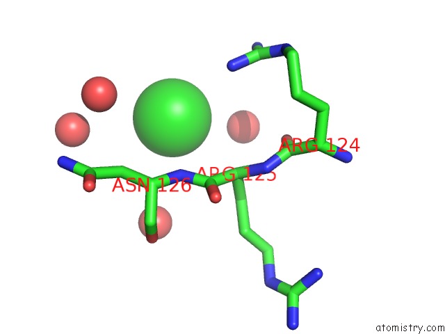
Mono view
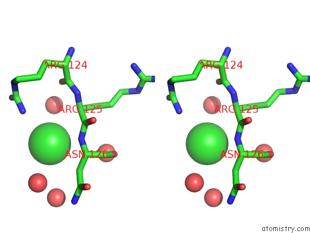
Stereo pair view
Mono view
Stereo pair view
A full contact list of Chlorine with other atoms in the Cl binding
site number 1 of Crystal Structure of the Sugar Recognizing Scf Ubiquitin Ligase at 1.7 Resolution within 5.0Å range:
|
Reference:
S.Vaijayanthimala,
D.Velmurugan,
T.Mizushima,
T.Yamane,
Y.Yoshida,
K.Tanaka.
Crystal Structure of the Sugar Recognizing Scf Ubiquitin Ligase at 1.7 Resolution To Be Published.
Page generated: Fri Jul 11 00:31:00 2025
Last articles
Na in 6ZTBNa in 6ZT3
Na in 6ZUK
Na in 6ZT2
Na in 6ZSN
Na in 6ZPL
Na in 6ZRA
Na in 6ZR6
Na in 6ZPQ
Na in 6ZO8