Chlorine »
PDB 2uxp-2v79 »
2uzc »
Chlorine in PDB 2uzc: Structure of Human PDLIM5 in Complex with the C-Terminal Peptide of Human Alpha-Actinin-1
Protein crystallography data
The structure of Structure of Human PDLIM5 in Complex with the C-Terminal Peptide of Human Alpha-Actinin-1, PDB code: 2uzc
was solved by
G.Bunkoczi,
J.Elkins,
E.Salah,
N.Burgess-Brown,
E.Papagrigoriou,
A.C.W.Pike,
A.Turnbull,
O.Gileadi,
F.Von Delft,
C.H.Arrowsmith,
A.Edwards,
M.Sundstrom,
J.Weigelt,
D.Doyle,
with X-Ray Crystallography technique. A brief refinement statistics is given in the table below:
| Resolution Low / High (Å) | 50.00 / 1.50 |
| Space group | P 1 21 1 |
| Cell size a, b, c (Å), α, β, γ (°) | 61.250, 36.474, 88.706, 90.00, 99.15, 90.00 |
| R / Rfree (%) | 16.3 / 21.5 |
Chlorine Binding Sites:
The binding sites of Chlorine atom in the Structure of Human PDLIM5 in Complex with the C-Terminal Peptide of Human Alpha-Actinin-1
(pdb code 2uzc). This binding sites where shown within
5.0 Angstroms radius around Chlorine atom.
In total 6 binding sites of Chlorine where determined in the Structure of Human PDLIM5 in Complex with the C-Terminal Peptide of Human Alpha-Actinin-1, PDB code: 2uzc:
Jump to Chlorine binding site number: 1; 2; 3; 4; 5; 6;
In total 6 binding sites of Chlorine where determined in the Structure of Human PDLIM5 in Complex with the C-Terminal Peptide of Human Alpha-Actinin-1, PDB code: 2uzc:
Jump to Chlorine binding site number: 1; 2; 3; 4; 5; 6;
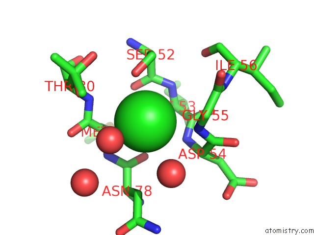
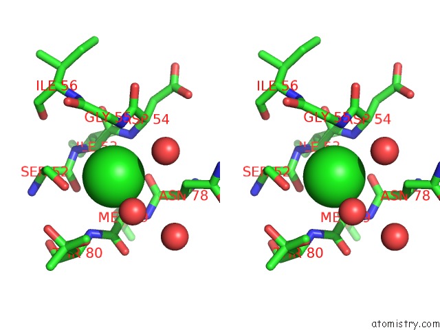
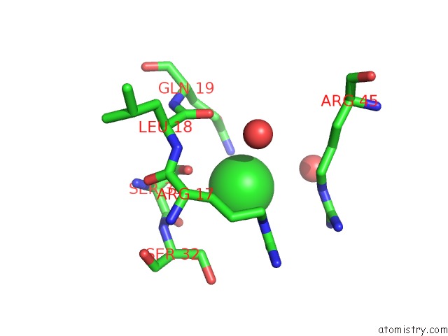
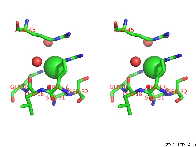
Chlorine binding site 1 out of 6 in 2uzc
Go back to
Chlorine binding site 1 out
of 6 in the Structure of Human PDLIM5 in Complex with the C-Terminal Peptide of Human Alpha-Actinin-1
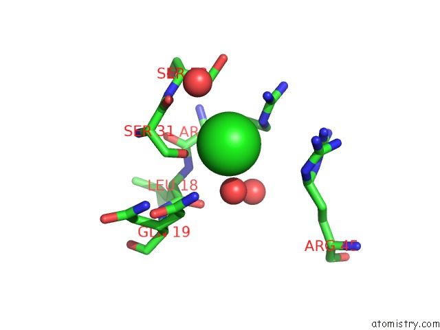
Mono view
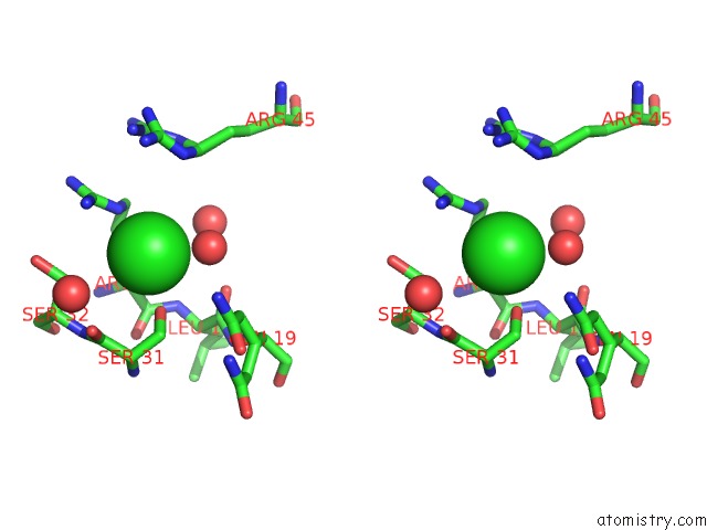
Stereo pair view
Mono view
Stereo pair view
A full contact list of Chlorine with other atoms in the Cl binding
site number 1 of Structure of Human PDLIM5 in Complex with the C-Terminal Peptide of Human Alpha-Actinin-1 within 5.0Å range:
|
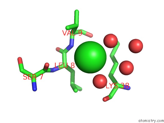
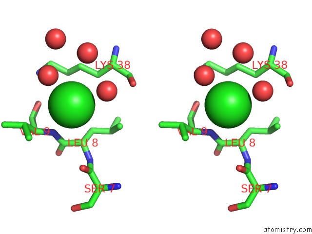
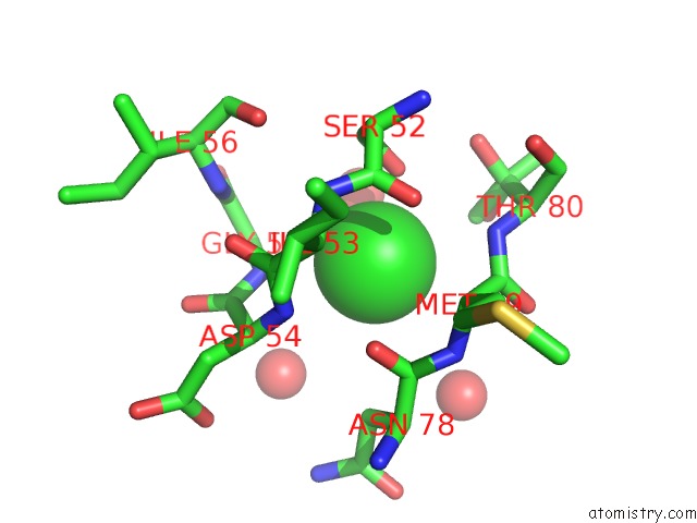
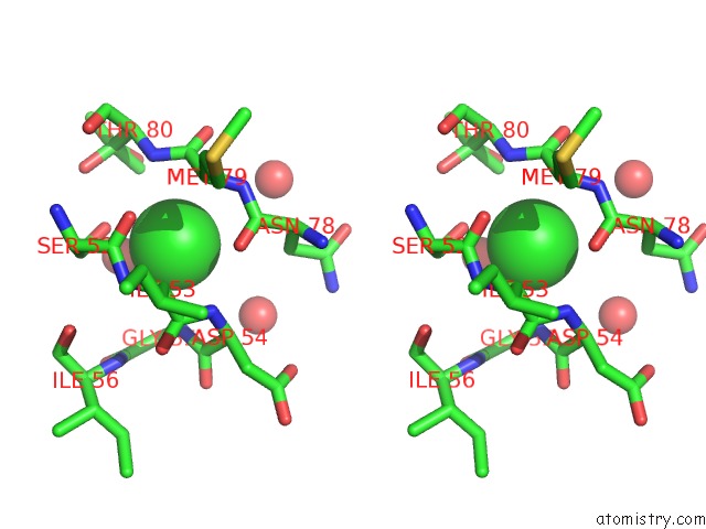
Chlorine binding site 2 out of 6 in 2uzc
Go back to
Chlorine binding site 2 out
of 6 in the Structure of Human PDLIM5 in Complex with the C-Terminal Peptide of Human Alpha-Actinin-1
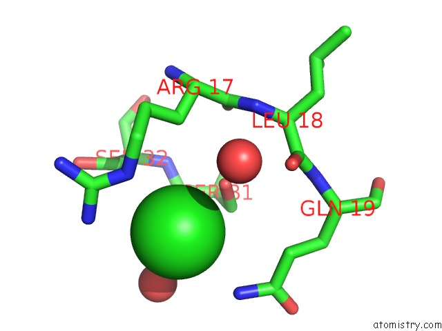
Mono view
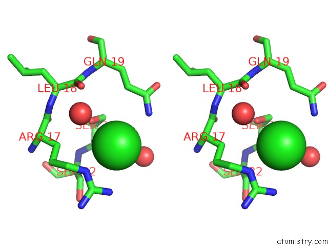
Stereo pair view
Mono view
Stereo pair view
A full contact list of Chlorine with other atoms in the Cl binding
site number 2 of Structure of Human PDLIM5 in Complex with the C-Terminal Peptide of Human Alpha-Actinin-1 within 5.0Å range:
|
Chlorine binding site 3 out of 6 in 2uzc
Go back to
Chlorine binding site 3 out
of 6 in the Structure of Human PDLIM5 in Complex with the C-Terminal Peptide of Human Alpha-Actinin-1
Mono view
Stereo pair view
Mono view
Stereo pair view
A full contact list of Chlorine with other atoms in the Cl binding
site number 3 of Structure of Human PDLIM5 in Complex with the C-Terminal Peptide of Human Alpha-Actinin-1 within 5.0Å range:
|
Chlorine binding site 4 out of 6 in 2uzc
Go back to
Chlorine binding site 4 out
of 6 in the Structure of Human PDLIM5 in Complex with the C-Terminal Peptide of Human Alpha-Actinin-1
Mono view
Stereo pair view
Mono view
Stereo pair view
A full contact list of Chlorine with other atoms in the Cl binding
site number 4 of Structure of Human PDLIM5 in Complex with the C-Terminal Peptide of Human Alpha-Actinin-1 within 5.0Å range:
|
Chlorine binding site 5 out of 6 in 2uzc
Go back to
Chlorine binding site 5 out
of 6 in the Structure of Human PDLIM5 in Complex with the C-Terminal Peptide of Human Alpha-Actinin-1
Mono view
Stereo pair view
Mono view
Stereo pair view
A full contact list of Chlorine with other atoms in the Cl binding
site number 5 of Structure of Human PDLIM5 in Complex with the C-Terminal Peptide of Human Alpha-Actinin-1 within 5.0Å range:
|
Chlorine binding site 6 out of 6 in 2uzc
Go back to
Chlorine binding site 6 out
of 6 in the Structure of Human PDLIM5 in Complex with the C-Terminal Peptide of Human Alpha-Actinin-1
Mono view
Stereo pair view
Mono view
Stereo pair view
A full contact list of Chlorine with other atoms in the Cl binding
site number 6 of Structure of Human PDLIM5 in Complex with the C-Terminal Peptide of Human Alpha-Actinin-1 within 5.0Å range:
|
Reference:
J.M.Elkins,
C.Gileadi,
L.Shrestha,
C.Phillips,
J.Wang,
J.R.Muniz,
D.A.Doyle.
Unusual Binding Interactions in Pdz Domain Crystal Structures Help Explain Binding Mechanisms. Protein Sci. V. 19 731 2010.
ISSN: ESSN 1469-896X
PubMed: 20120020
DOI: 10.1002/PRO.349
Page generated: Fri Jul 11 00:35:26 2025
ISSN: ESSN 1469-896X
PubMed: 20120020
DOI: 10.1002/PRO.349
Last articles
Fe in 2YXOFe in 2YRS
Fe in 2YXC
Fe in 2YNM
Fe in 2YVJ
Fe in 2YP1
Fe in 2YU2
Fe in 2YU1
Fe in 2YQB
Fe in 2YOO