Chlorine »
PDB 2vg2-2vo5 »
2vli »
Chlorine in PDB 2vli: Structure of Deinococcus Radiodurans Tunicamycin Resistance Protein
Protein crystallography data
The structure of Structure of Deinococcus Radiodurans Tunicamycin Resistance Protein, PDB code: 2vli
was solved by
S.Macedo,
U.Kapp,
I.Leiros,
D.R.Hall,
E.Mitchell,
with X-Ray Crystallography technique. A brief refinement statistics is given in the table below:
| Resolution Low / High (Å) | 22.90 / 1.95 |
| Space group | C 2 2 21 |
| Cell size a, b, c (Å), α, β, γ (°) | 81.360, 118.160, 81.110, 90.00, 90.00, 90.00 |
| R / Rfree (%) | 17.4 / 21.4 |
Other elements in 2vli:
The structure of Structure of Deinococcus Radiodurans Tunicamycin Resistance Protein also contains other interesting chemical elements:
| Cadmium | (Cd) | 1 atom |
Chlorine Binding Sites:
The binding sites of Chlorine atom in the Structure of Deinococcus Radiodurans Tunicamycin Resistance Protein
(pdb code 2vli). This binding sites where shown within
5.0 Angstroms radius around Chlorine atom.
In total only one binding site of Chlorine was determined in the Structure of Deinococcus Radiodurans Tunicamycin Resistance Protein, PDB code: 2vli:
In total only one binding site of Chlorine was determined in the Structure of Deinococcus Radiodurans Tunicamycin Resistance Protein, PDB code: 2vli:
Chlorine binding site 1 out of 1 in 2vli
Go back to
Chlorine binding site 1 out
of 1 in the Structure of Deinococcus Radiodurans Tunicamycin Resistance Protein
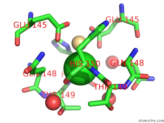
Mono view
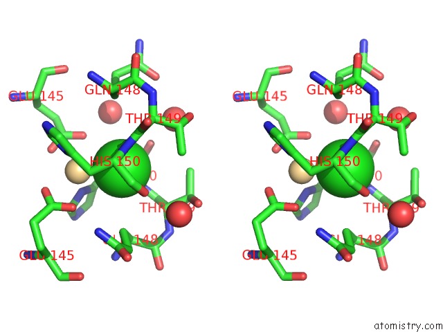
Stereo pair view
Mono view
Stereo pair view
A full contact list of Chlorine with other atoms in the Cl binding
site number 1 of Structure of Deinococcus Radiodurans Tunicamycin Resistance Protein within 5.0Å range:
|
Reference:
U.Kapp,
S.Macedo,
D.R.Hall,
I.Leiros,
S.M.Mcsweeney,
E.Mitchell.
Structure of Deinococcus Radiodurans Tunicamycin-Resistance Protein (Tmrd), A Phosphotransferase. Acta Crystallogr.,Sect.F V. 64 479 2008.
ISSN: ESSN 1744-3091
PubMed: 18540055
DOI: 10.1107/S1744309108011822
Page generated: Fri Jul 11 00:52:59 2025
ISSN: ESSN 1744-3091
PubMed: 18540055
DOI: 10.1107/S1744309108011822
Last articles
Mn in 9LJUMn in 9LJW
Mn in 9LJS
Mn in 9LJR
Mn in 9LJT
Mn in 9LJV
Mg in 9UA2
Mg in 9R96
Mg in 9VM1
Mg in 9P01