Chlorine »
PDB 2wzd-2x8b »
2x5k »
Chlorine in PDB 2x5k: Structure of An Active Site Mutant of the D-Erythrose-4-Phosphate Dehydrogenase From E. Coli
Enzymatic activity of Structure of An Active Site Mutant of the D-Erythrose-4-Phosphate Dehydrogenase From E. Coli
All present enzymatic activity of Structure of An Active Site Mutant of the D-Erythrose-4-Phosphate Dehydrogenase From E. Coli:
1.2.1.72;
1.2.1.72;
Protein crystallography data
The structure of Structure of An Active Site Mutant of the D-Erythrose-4-Phosphate Dehydrogenase From E. Coli, PDB code: 2x5k
was solved by
S.Moniot,
C.Didierjean,
S.Boschi-Muller,
G.Branlant,
C.Corbier,
with X-Ray Crystallography technique. A brief refinement statistics is given in the table below:
| Resolution Low / High (Å) | 55.97 / 2.37 |
| Space group | P 41 21 2 |
| Cell size a, b, c (Å), α, β, γ (°) | 134.810, 134.810, 246.110, 90.00, 90.00, 90.00 |
| R / Rfree (%) | 16.444 / 20.699 |
Chlorine Binding Sites:
The binding sites of Chlorine atom in the Structure of An Active Site Mutant of the D-Erythrose-4-Phosphate Dehydrogenase From E. Coli
(pdb code 2x5k). This binding sites where shown within
5.0 Angstroms radius around Chlorine atom.
In total 3 binding sites of Chlorine where determined in the Structure of An Active Site Mutant of the D-Erythrose-4-Phosphate Dehydrogenase From E. Coli, PDB code: 2x5k:
Jump to Chlorine binding site number: 1; 2; 3;
In total 3 binding sites of Chlorine where determined in the Structure of An Active Site Mutant of the D-Erythrose-4-Phosphate Dehydrogenase From E. Coli, PDB code: 2x5k:
Jump to Chlorine binding site number: 1; 2; 3;
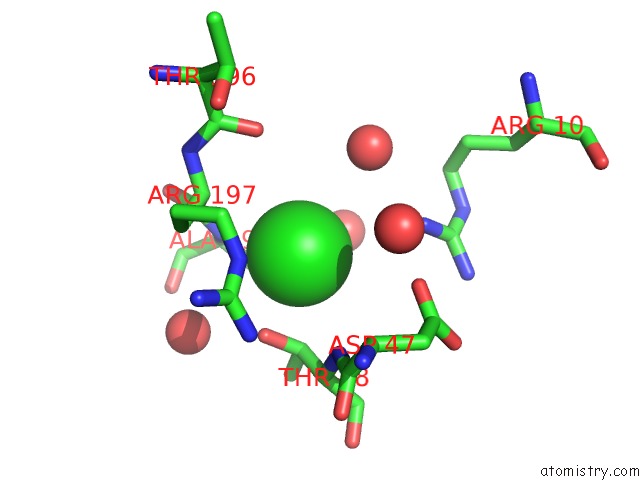
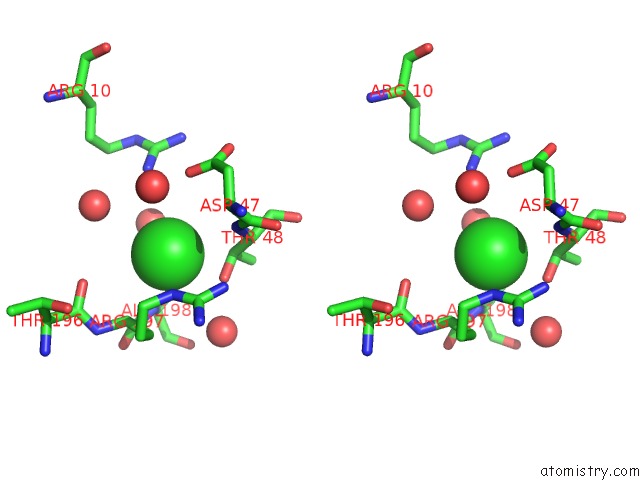
Chlorine binding site 1 out of 3 in 2x5k
Go back to
Chlorine binding site 1 out
of 3 in the Structure of An Active Site Mutant of the D-Erythrose-4-Phosphate Dehydrogenase From E. Coli
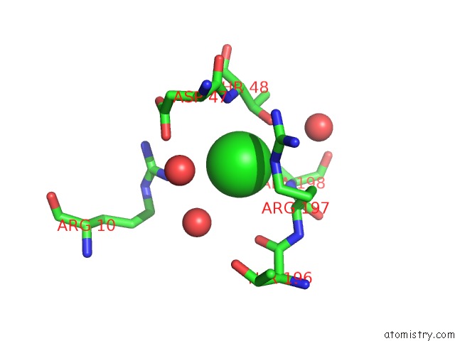
Mono view
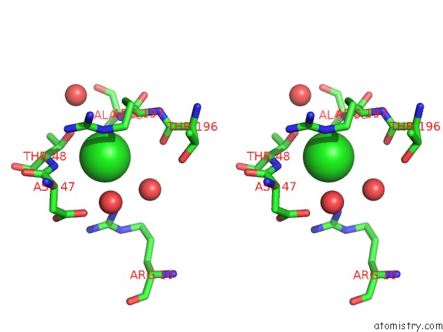
Stereo pair view
Mono view
Stereo pair view
A full contact list of Chlorine with other atoms in the Cl binding
site number 1 of Structure of An Active Site Mutant of the D-Erythrose-4-Phosphate Dehydrogenase From E. Coli within 5.0Å range:
|
Chlorine binding site 2 out of 3 in 2x5k
Go back to
Chlorine binding site 2 out
of 3 in the Structure of An Active Site Mutant of the D-Erythrose-4-Phosphate Dehydrogenase From E. Coli
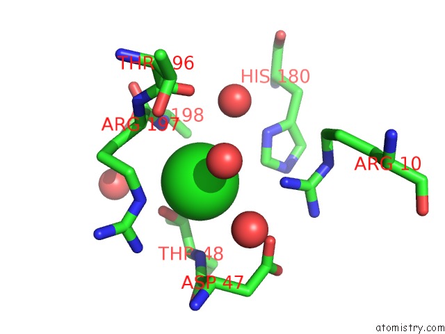
Mono view
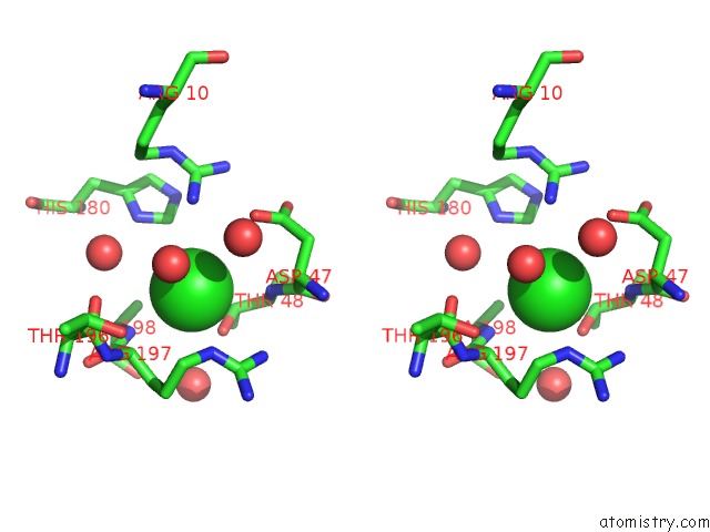
Stereo pair view
Mono view
Stereo pair view
A full contact list of Chlorine with other atoms in the Cl binding
site number 2 of Structure of An Active Site Mutant of the D-Erythrose-4-Phosphate Dehydrogenase From E. Coli within 5.0Å range:
|
Chlorine binding site 3 out of 3 in 2x5k
Go back to
Chlorine binding site 3 out
of 3 in the Structure of An Active Site Mutant of the D-Erythrose-4-Phosphate Dehydrogenase From E. Coli
Mono view
Stereo pair view
Mono view
Stereo pair view
A full contact list of Chlorine with other atoms in the Cl binding
site number 3 of Structure of An Active Site Mutant of the D-Erythrose-4-Phosphate Dehydrogenase From E. Coli within 5.0Å range:
|
Reference:
S.Moniot,
C.Didierjean,
S.Boschi-Muller,
G.Branlant,
C.Corbier.
Structural Characterization of Erythrose-4- Phosphate Dehydrogenase From Escherichia Coli: Peculiar Features When Compared to Phosphorylating Gapdhs To Be Published.
Page generated: Fri Jul 11 01:46:20 2025
Last articles
Na in 4TR8Na in 4TPU
Na in 4TR6
Na in 4TOG
Na in 4TOF
Na in 4TPR
Na in 4TMX
Na in 4TMW
Na in 4TME
Na in 4TMV