Chlorine »
PDB 3akg-3avh »
3ap9 »
Chlorine in PDB 3ap9: Crystal Structure of the Galectin-8 N-Terminal Carbohydrate Recognition Domain in Complex with Lacto-N-Fucopentaose III
Protein crystallography data
The structure of Crystal Structure of the Galectin-8 N-Terminal Carbohydrate Recognition Domain in Complex with Lacto-N-Fucopentaose III, PDB code: 3ap9
was solved by
T.Matsuzaka,
H.Ideo,
K.Yamashita,
T.Nonaka,
with X-Ray Crystallography technique. A brief refinement statistics is given in the table below:
| Resolution Low / High (Å) | 24.98 / 1.33 |
| Space group | P 21 21 21 |
| Cell size a, b, c (Å), α, β, γ (°) | 46.419, 49.952, 70.207, 90.00, 90.00, 90.00 |
| R / Rfree (%) | 15.7 / 17.8 |
Chlorine Binding Sites:
The binding sites of Chlorine atom in the Crystal Structure of the Galectin-8 N-Terminal Carbohydrate Recognition Domain in Complex with Lacto-N-Fucopentaose III
(pdb code 3ap9). This binding sites where shown within
5.0 Angstroms radius around Chlorine atom.
In total 3 binding sites of Chlorine where determined in the Crystal Structure of the Galectin-8 N-Terminal Carbohydrate Recognition Domain in Complex with Lacto-N-Fucopentaose III, PDB code: 3ap9:
Jump to Chlorine binding site number: 1; 2; 3;
In total 3 binding sites of Chlorine where determined in the Crystal Structure of the Galectin-8 N-Terminal Carbohydrate Recognition Domain in Complex with Lacto-N-Fucopentaose III, PDB code: 3ap9:
Jump to Chlorine binding site number: 1; 2; 3;
Chlorine binding site 1 out of 3 in 3ap9
Go back to
Chlorine binding site 1 out
of 3 in the Crystal Structure of the Galectin-8 N-Terminal Carbohydrate Recognition Domain in Complex with Lacto-N-Fucopentaose III
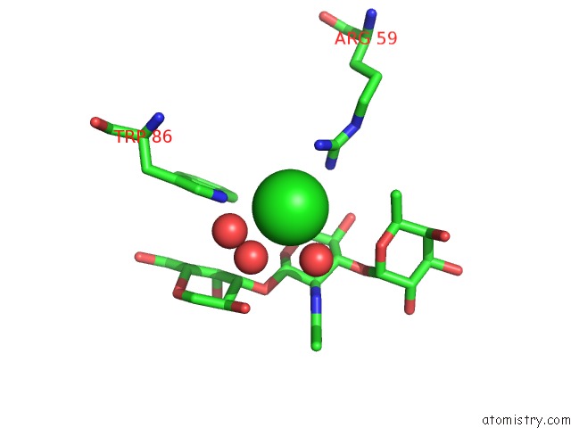
Mono view

Stereo pair view
Mono view
Stereo pair view
A full contact list of Chlorine with other atoms in the Cl binding
site number 1 of Crystal Structure of the Galectin-8 N-Terminal Carbohydrate Recognition Domain in Complex with Lacto-N-Fucopentaose III within 5.0Å range:
|
Chlorine binding site 2 out of 3 in 3ap9
Go back to
Chlorine binding site 2 out
of 3 in the Crystal Structure of the Galectin-8 N-Terminal Carbohydrate Recognition Domain in Complex with Lacto-N-Fucopentaose III

Mono view

Stereo pair view
Mono view
Stereo pair view
A full contact list of Chlorine with other atoms in the Cl binding
site number 2 of Crystal Structure of the Galectin-8 N-Terminal Carbohydrate Recognition Domain in Complex with Lacto-N-Fucopentaose III within 5.0Å range:
|
Chlorine binding site 3 out of 3 in 3ap9
Go back to
Chlorine binding site 3 out
of 3 in the Crystal Structure of the Galectin-8 N-Terminal Carbohydrate Recognition Domain in Complex with Lacto-N-Fucopentaose III

Mono view
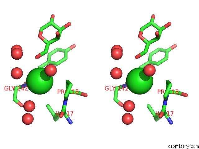
Stereo pair view
Mono view
Stereo pair view
A full contact list of Chlorine with other atoms in the Cl binding
site number 3 of Crystal Structure of the Galectin-8 N-Terminal Carbohydrate Recognition Domain in Complex with Lacto-N-Fucopentaose III within 5.0Å range:
|
Reference:
H.Ideo,
T.Matsuzaka,
T.Nonaka,
A.Seko,
K.Yamashita.
Galectin-8-N-Domain Recognition Mechanism For Sialylated and Sulfated Glycans J.Biol.Chem. V. 286 11346 2011.
ISSN: ISSN 0021-9258
PubMed: 21288902
DOI: 10.1074/JBC.M110.195925
Page generated: Fri Jul 11 03:09:32 2025
ISSN: ISSN 0021-9258
PubMed: 21288902
DOI: 10.1074/JBC.M110.195925
Last articles
Na in 1ZJNNa in 1ZL0
Na in 1ZJM
Na in 1ZKO
Na in 1ZH8
Na in 1ZFA
Na in 1ZF9
Na in 1ZF7
Na in 1ZF6
Na in 1ZDN