Chlorine »
PDB 3b0x-3bft »
3be6 »
Chlorine in PDB 3be6: Crystal Structure of Fite (Crystal Form 2), A Group III Periplasmic Siderophore Binding Protein
Protein crystallography data
The structure of Crystal Structure of Fite (Crystal Form 2), A Group III Periplasmic Siderophore Binding Protein, PDB code: 3be6
was solved by
R.Shi,
A.Matte,
M.Cygler,
Montreal-Kingston Bacterial Structuralgenomics Initiative (Bsgi),
with X-Ray Crystallography technique. A brief refinement statistics is given in the table below:
| Resolution Low / High (Å) | 49.45 / 1.82 |
| Space group | P 21 21 21 |
| Cell size a, b, c (Å), α, β, γ (°) | 50.815, 109.120, 221.992, 90.00, 90.00, 90.00 |
| R / Rfree (%) | 18.8 / 21.9 |
Other elements in 3be6:
The structure of Crystal Structure of Fite (Crystal Form 2), A Group III Periplasmic Siderophore Binding Protein also contains other interesting chemical elements:
| Magnesium | (Mg) | 1 atom |
Chlorine Binding Sites:
The binding sites of Chlorine atom in the Crystal Structure of Fite (Crystal Form 2), A Group III Periplasmic Siderophore Binding Protein
(pdb code 3be6). This binding sites where shown within
5.0 Angstroms radius around Chlorine atom.
In total 7 binding sites of Chlorine where determined in the Crystal Structure of Fite (Crystal Form 2), A Group III Periplasmic Siderophore Binding Protein, PDB code: 3be6:
Jump to Chlorine binding site number: 1; 2; 3; 4; 5; 6; 7;
In total 7 binding sites of Chlorine where determined in the Crystal Structure of Fite (Crystal Form 2), A Group III Periplasmic Siderophore Binding Protein, PDB code: 3be6:
Jump to Chlorine binding site number: 1; 2; 3; 4; 5; 6; 7;
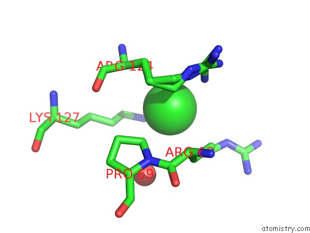
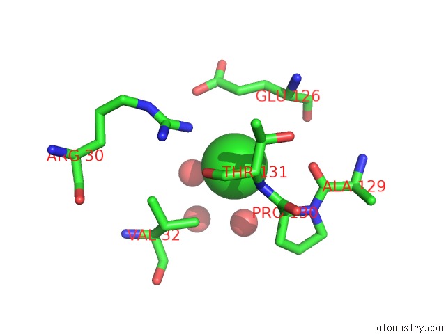
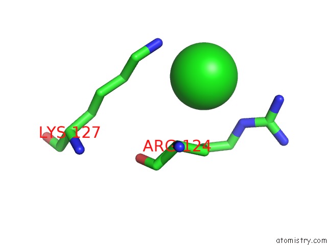
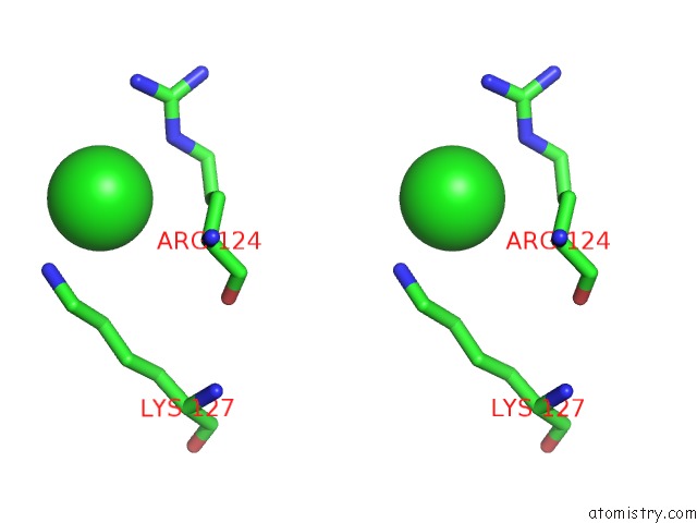
Chlorine binding site 1 out of 7 in 3be6
Go back to
Chlorine binding site 1 out
of 7 in the Crystal Structure of Fite (Crystal Form 2), A Group III Periplasmic Siderophore Binding Protein
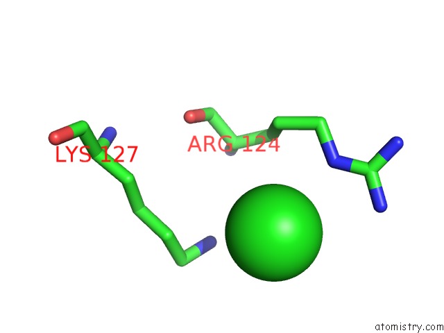
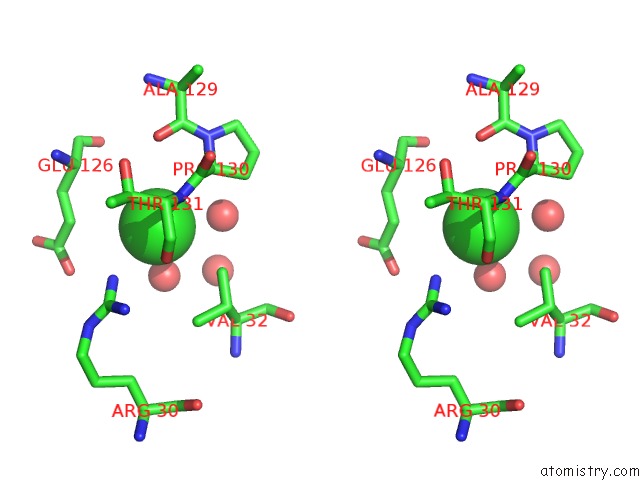
Mono view
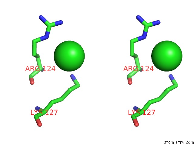
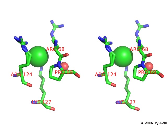
Stereo pair view
Mono view
Stereo pair view
A full contact list of Chlorine with other atoms in the Cl binding
site number 1 of Crystal Structure of Fite (Crystal Form 2), A Group III Periplasmic Siderophore Binding Protein within 5.0Å range:
|
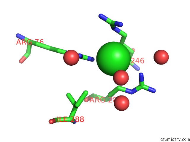
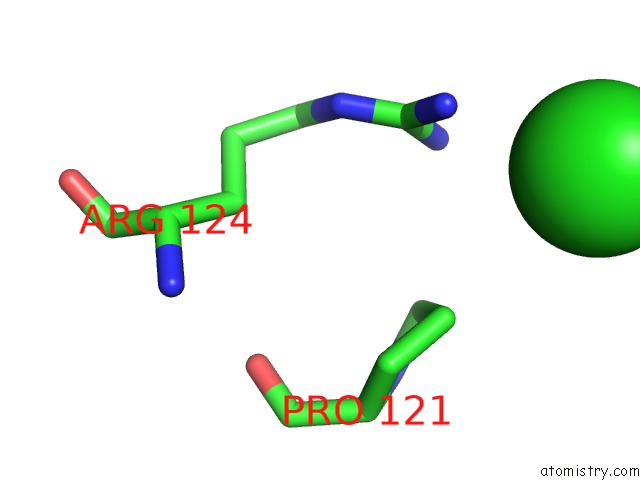
Chlorine binding site 2 out of 7 in 3be6
Go back to
Chlorine binding site 2 out
of 7 in the Crystal Structure of Fite (Crystal Form 2), A Group III Periplasmic Siderophore Binding Protein
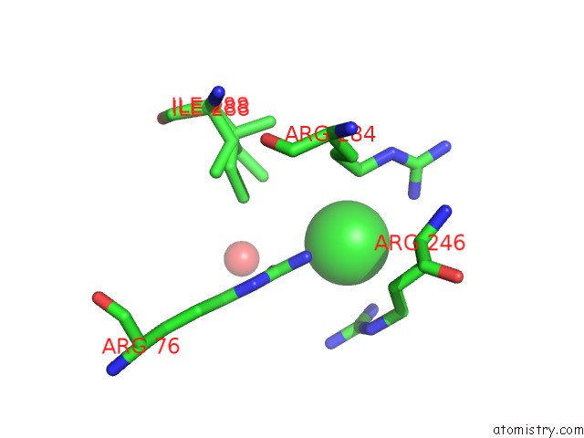
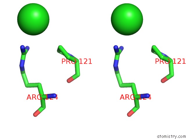
Mono view
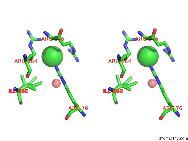
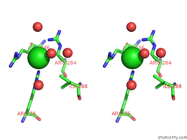
Stereo pair view
Mono view
Stereo pair view
A full contact list of Chlorine with other atoms in the Cl binding
site number 2 of Crystal Structure of Fite (Crystal Form 2), A Group III Periplasmic Siderophore Binding Protein within 5.0Å range:
|
Chlorine binding site 3 out of 7 in 3be6
Go back to
Chlorine binding site 3 out
of 7 in the Crystal Structure of Fite (Crystal Form 2), A Group III Periplasmic Siderophore Binding Protein
Mono view
Stereo pair view
Mono view
Stereo pair view
A full contact list of Chlorine with other atoms in the Cl binding
site number 3 of Crystal Structure of Fite (Crystal Form 2), A Group III Periplasmic Siderophore Binding Protein within 5.0Å range:
|
Chlorine binding site 4 out of 7 in 3be6
Go back to
Chlorine binding site 4 out
of 7 in the Crystal Structure of Fite (Crystal Form 2), A Group III Periplasmic Siderophore Binding Protein
Mono view
Stereo pair view
Mono view
Stereo pair view
A full contact list of Chlorine with other atoms in the Cl binding
site number 4 of Crystal Structure of Fite (Crystal Form 2), A Group III Periplasmic Siderophore Binding Protein within 5.0Å range:
|
Chlorine binding site 5 out of 7 in 3be6
Go back to
Chlorine binding site 5 out
of 7 in the Crystal Structure of Fite (Crystal Form 2), A Group III Periplasmic Siderophore Binding Protein
Mono view
Stereo pair view
Mono view
Stereo pair view
A full contact list of Chlorine with other atoms in the Cl binding
site number 5 of Crystal Structure of Fite (Crystal Form 2), A Group III Periplasmic Siderophore Binding Protein within 5.0Å range:
|
Chlorine binding site 6 out of 7 in 3be6
Go back to
Chlorine binding site 6 out
of 7 in the Crystal Structure of Fite (Crystal Form 2), A Group III Periplasmic Siderophore Binding Protein
Mono view
Stereo pair view
Mono view
Stereo pair view
A full contact list of Chlorine with other atoms in the Cl binding
site number 6 of Crystal Structure of Fite (Crystal Form 2), A Group III Periplasmic Siderophore Binding Protein within 5.0Å range:
|
Chlorine binding site 7 out of 7 in 3be6
Go back to
Chlorine binding site 7 out
of 7 in the Crystal Structure of Fite (Crystal Form 2), A Group III Periplasmic Siderophore Binding Protein
Mono view
Stereo pair view
Mono view
Stereo pair view
A full contact list of Chlorine with other atoms in the Cl binding
site number 7 of Crystal Structure of Fite (Crystal Form 2), A Group III Periplasmic Siderophore Binding Protein within 5.0Å range:
|
Reference:
R.Shi,
A.Proteau,
J.Wagner,
Q.Cui,
E.O.Purisima,
A.Matte,
M.Cygler.
Trapping Open and Closed Forms of Fite-A Group III Periplasmic Binding Protein. Proteins V. 75 598 2008.
ISSN: ISSN 0887-3585
PubMed: 19004000
DOI: 10.1002/PROT.22272
Page generated: Fri Jul 11 03:25:35 2025
ISSN: ISSN 0887-3585
PubMed: 19004000
DOI: 10.1002/PROT.22272
Last articles
Mg in 6JUOMg in 6JUN
Mg in 6JUM
Mg in 6JUL
Mg in 6JTA
Mg in 6JTG
Mg in 6JT9
Mg in 6JT8
Mg in 6JT7
Mg in 6JT2