Chlorine »
PDB 3ccq-3ck1 »
3cd3 »
Chlorine in PDB 3cd3: Crystal Structure of Phosphorylated Human Feline Sarcoma Viral Oncogene Homologue (V-Fes) in Complex with Staurosporine and A Consensus Peptide
Enzymatic activity of Crystal Structure of Phosphorylated Human Feline Sarcoma Viral Oncogene Homologue (V-Fes) in Complex with Staurosporine and A Consensus Peptide
All present enzymatic activity of Crystal Structure of Phosphorylated Human Feline Sarcoma Viral Oncogene Homologue (V-Fes) in Complex with Staurosporine and A Consensus Peptide:
2.7.10.2;
2.7.10.2;
Protein crystallography data
The structure of Crystal Structure of Phosphorylated Human Feline Sarcoma Viral Oncogene Homologue (V-Fes) in Complex with Staurosporine and A Consensus Peptide, PDB code: 3cd3
was solved by
P.Filippakopoulos,
E.Salah,
C.Cooper,
S.S.Picaud,
J.M.Elkins,
F.Von Delft,
C.H.Arrowsmith,
A.M.Edwards,
J.Weigelt,
C.Bountra,
S.Knapp,
Structuralgenomics Consortium (Sgc),
with X-Ray Crystallography technique. A brief refinement statistics is given in the table below:
| Resolution Low / High (Å) | 15.35 / 1.98 |
| Space group | P 21 21 21 |
| Cell size a, b, c (Å), α, β, γ (°) | 35.451, 76.905, 150.632, 90.00, 90.00, 90.00 |
| R / Rfree (%) | 18.5 / 24.7 |
Chlorine Binding Sites:
The binding sites of Chlorine atom in the Crystal Structure of Phosphorylated Human Feline Sarcoma Viral Oncogene Homologue (V-Fes) in Complex with Staurosporine and A Consensus Peptide
(pdb code 3cd3). This binding sites where shown within
5.0 Angstroms radius around Chlorine atom.
In total 2 binding sites of Chlorine where determined in the Crystal Structure of Phosphorylated Human Feline Sarcoma Viral Oncogene Homologue (V-Fes) in Complex with Staurosporine and A Consensus Peptide, PDB code: 3cd3:
Jump to Chlorine binding site number: 1; 2;
In total 2 binding sites of Chlorine where determined in the Crystal Structure of Phosphorylated Human Feline Sarcoma Viral Oncogene Homologue (V-Fes) in Complex with Staurosporine and A Consensus Peptide, PDB code: 3cd3:
Jump to Chlorine binding site number: 1; 2;
Chlorine binding site 1 out of 2 in 3cd3
Go back to
Chlorine binding site 1 out
of 2 in the Crystal Structure of Phosphorylated Human Feline Sarcoma Viral Oncogene Homologue (V-Fes) in Complex with Staurosporine and A Consensus Peptide
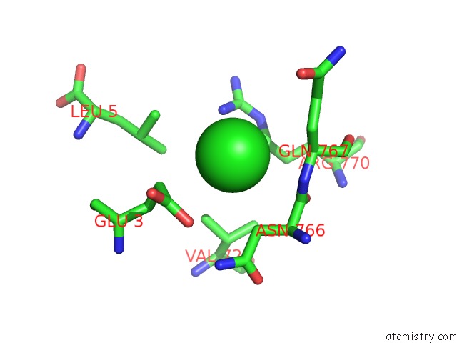
Mono view
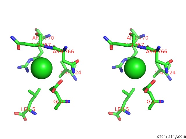
Stereo pair view
Mono view
Stereo pair view
A full contact list of Chlorine with other atoms in the Cl binding
site number 1 of Crystal Structure of Phosphorylated Human Feline Sarcoma Viral Oncogene Homologue (V-Fes) in Complex with Staurosporine and A Consensus Peptide within 5.0Å range:
|
Chlorine binding site 2 out of 2 in 3cd3
Go back to
Chlorine binding site 2 out
of 2 in the Crystal Structure of Phosphorylated Human Feline Sarcoma Viral Oncogene Homologue (V-Fes) in Complex with Staurosporine and A Consensus Peptide
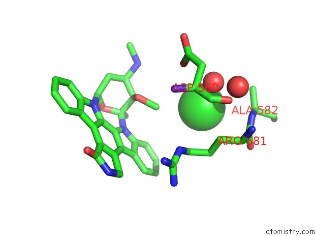
Mono view
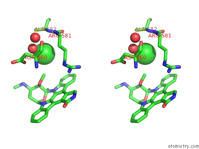
Stereo pair view
Mono view
Stereo pair view
A full contact list of Chlorine with other atoms in the Cl binding
site number 2 of Crystal Structure of Phosphorylated Human Feline Sarcoma Viral Oncogene Homologue (V-Fes) in Complex with Staurosporine and A Consensus Peptide within 5.0Å range:
|
Reference:
P.Filippakopoulos,
M.Kofler,
O.Hantschel,
G.D.Gish,
F.Grebien,
E.Salah,
P.Neudecker,
L.E.Kay,
B.E.Turk,
G.Superti-Furga,
T.Pawson,
S.Knapp.
Structural Coupling of SH2-Kinase Domains Links Fes and Abl Substrate Recognition and Kinase Activation Cell(Cambridge,Mass.) V. 134 793 2008.
ISSN: ISSN 0092-8674
PubMed: 18775312
DOI: 10.1016/J.CELL.2008.07.047
Page generated: Fri Jul 11 03:56:56 2025
ISSN: ISSN 0092-8674
PubMed: 18775312
DOI: 10.1016/J.CELL.2008.07.047
Last articles
I in 8E59I in 8E58
I in 8E57
I in 8DC1
I in 8E56
I in 8DXL
I in 8DV3
I in 8DC5
I in 8DGT
I in 8D8W