Chlorine »
PDB 3ck2-3csw »
3cmb »
Chlorine in PDB 3cmb: Crystal Structure of Acetoacetate Decarboxylase (YP_001047042.1) From Methanoculleus Marisnigri JR1 at 1.60 A Resolution
Protein crystallography data
The structure of Crystal Structure of Acetoacetate Decarboxylase (YP_001047042.1) From Methanoculleus Marisnigri JR1 at 1.60 A Resolution, PDB code: 3cmb
was solved by
Joint Center For Structural Genomics (Jcsg),
with X-Ray Crystallography technique. A brief refinement statistics is given in the table below:
| Resolution Low / High (Å) | 29.39 / 1.60 |
| Space group | I 2 2 2 |
| Cell size a, b, c (Å), α, β, γ (°) | 129.340, 136.710, 168.120, 90.00, 90.00, 90.00 |
| R / Rfree (%) | 22.8 / 25.9 |
Other elements in 3cmb:
The structure of Crystal Structure of Acetoacetate Decarboxylase (YP_001047042.1) From Methanoculleus Marisnigri JR1 at 1.60 A Resolution also contains other interesting chemical elements:
| Sodium | (Na) | 12 atoms |
Chlorine Binding Sites:
The binding sites of Chlorine atom in the Crystal Structure of Acetoacetate Decarboxylase (YP_001047042.1) From Methanoculleus Marisnigri JR1 at 1.60 A Resolution
(pdb code 3cmb). This binding sites where shown within
5.0 Angstroms radius around Chlorine atom.
In total 4 binding sites of Chlorine where determined in the Crystal Structure of Acetoacetate Decarboxylase (YP_001047042.1) From Methanoculleus Marisnigri JR1 at 1.60 A Resolution, PDB code: 3cmb:
Jump to Chlorine binding site number: 1; 2; 3; 4;
In total 4 binding sites of Chlorine where determined in the Crystal Structure of Acetoacetate Decarboxylase (YP_001047042.1) From Methanoculleus Marisnigri JR1 at 1.60 A Resolution, PDB code: 3cmb:
Jump to Chlorine binding site number: 1; 2; 3; 4;
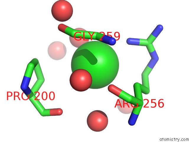
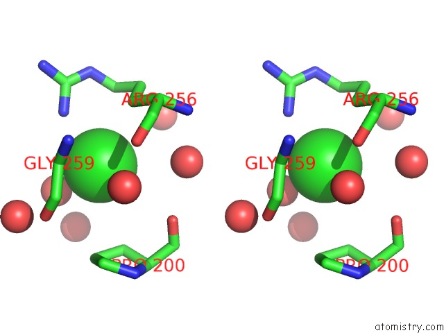
Chlorine binding site 1 out of 4 in 3cmb
Go back to
Chlorine binding site 1 out
of 4 in the Crystal Structure of Acetoacetate Decarboxylase (YP_001047042.1) From Methanoculleus Marisnigri JR1 at 1.60 A Resolution
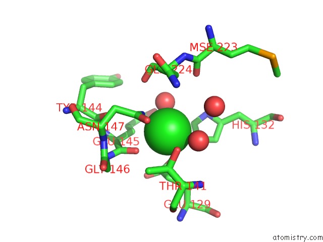
Mono view
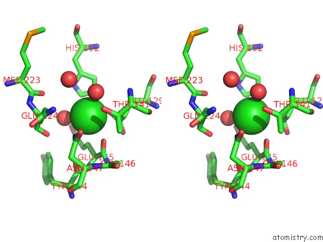
Stereo pair view
Mono view
Stereo pair view
A full contact list of Chlorine with other atoms in the Cl binding
site number 1 of Crystal Structure of Acetoacetate Decarboxylase (YP_001047042.1) From Methanoculleus Marisnigri JR1 at 1.60 A Resolution within 5.0Å range:
|
Chlorine binding site 2 out of 4 in 3cmb
Go back to
Chlorine binding site 2 out
of 4 in the Crystal Structure of Acetoacetate Decarboxylase (YP_001047042.1) From Methanoculleus Marisnigri JR1 at 1.60 A Resolution
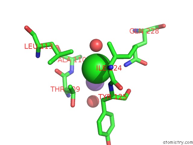
Mono view
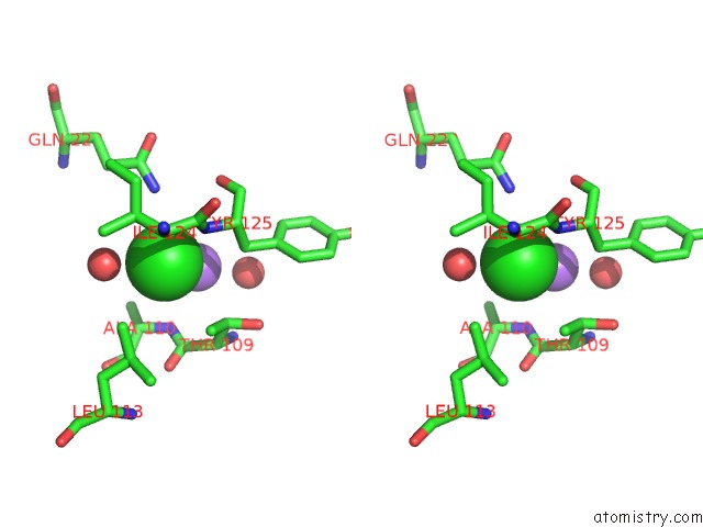
Stereo pair view
Mono view
Stereo pair view
A full contact list of Chlorine with other atoms in the Cl binding
site number 2 of Crystal Structure of Acetoacetate Decarboxylase (YP_001047042.1) From Methanoculleus Marisnigri JR1 at 1.60 A Resolution within 5.0Å range:
|
Chlorine binding site 3 out of 4 in 3cmb
Go back to
Chlorine binding site 3 out
of 4 in the Crystal Structure of Acetoacetate Decarboxylase (YP_001047042.1) From Methanoculleus Marisnigri JR1 at 1.60 A Resolution
Mono view
Stereo pair view
Mono view
Stereo pair view
A full contact list of Chlorine with other atoms in the Cl binding
site number 3 of Crystal Structure of Acetoacetate Decarboxylase (YP_001047042.1) From Methanoculleus Marisnigri JR1 at 1.60 A Resolution within 5.0Å range:
|
Chlorine binding site 4 out of 4 in 3cmb
Go back to
Chlorine binding site 4 out
of 4 in the Crystal Structure of Acetoacetate Decarboxylase (YP_001047042.1) From Methanoculleus Marisnigri JR1 at 1.60 A Resolution

Mono view
Stereo pair view
Mono view
Stereo pair view
A full contact list of Chlorine with other atoms in the Cl binding
site number 4 of Crystal Structure of Acetoacetate Decarboxylase (YP_001047042.1) From Methanoculleus Marisnigri JR1 at 1.60 A Resolution within 5.0Å range:
|
Reference:
Joint Center For Structural Genomics (Jcsg),
Joint Center For Structural Genomics (Jcsg).
N/A N/A.
Page generated: Fri Jul 11 04:07:03 2025
Last articles
Mg in 3C9UMg in 3CBT
Mg in 3CBQ
Mg in 3CBG
Mg in 3CBE
Mg in 3C9T
Mg in 3CB9
Mg in 3CAW
Mg in 3CA9
Mg in 3C5P