Chlorine »
PDB 3dzt-3ec8 »
3e7b »
Chlorine in PDB 3e7b: Crystal Structure of Protein Phosphatase-1 Bound to the Natural Toxin Inhibitor Tautomycin
Enzymatic activity of Crystal Structure of Protein Phosphatase-1 Bound to the Natural Toxin Inhibitor Tautomycin
All present enzymatic activity of Crystal Structure of Protein Phosphatase-1 Bound to the Natural Toxin Inhibitor Tautomycin:
3.1.3.16;
3.1.3.16;
Protein crystallography data
The structure of Crystal Structure of Protein Phosphatase-1 Bound to the Natural Toxin Inhibitor Tautomycin, PDB code: 3e7b
was solved by
M.S.Kelker,
R.Page,
W.Peti,
with X-Ray Crystallography technique. A brief refinement statistics is given in the table below:
| Resolution Low / High (Å) | 19.63 / 1.70 |
| Space group | P 21 21 21 |
| Cell size a, b, c (Å), α, β, γ (°) | 65.760, 78.519, 130.764, 90.00, 90.00, 90.00 |
| R / Rfree (%) | 15.3 / 17.5 |
Other elements in 3e7b:
The structure of Crystal Structure of Protein Phosphatase-1 Bound to the Natural Toxin Inhibitor Tautomycin also contains other interesting chemical elements:
| Manganese | (Mn) | 4 atoms |
| Sodium | (Na) | 1 atom |
Chlorine Binding Sites:
The binding sites of Chlorine atom in the Crystal Structure of Protein Phosphatase-1 Bound to the Natural Toxin Inhibitor Tautomycin
(pdb code 3e7b). This binding sites where shown within
5.0 Angstroms radius around Chlorine atom.
In total only one binding site of Chlorine was determined in the Crystal Structure of Protein Phosphatase-1 Bound to the Natural Toxin Inhibitor Tautomycin, PDB code: 3e7b:
In total only one binding site of Chlorine was determined in the Crystal Structure of Protein Phosphatase-1 Bound to the Natural Toxin Inhibitor Tautomycin, PDB code: 3e7b:
Chlorine binding site 1 out of 1 in 3e7b
Go back to
Chlorine binding site 1 out
of 1 in the Crystal Structure of Protein Phosphatase-1 Bound to the Natural Toxin Inhibitor Tautomycin
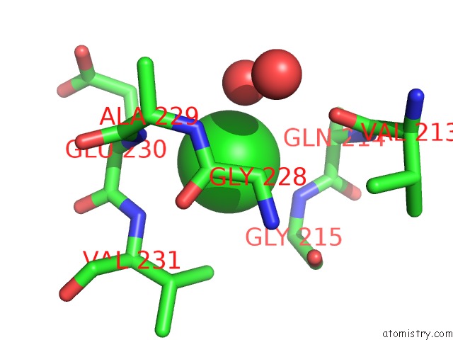
Mono view
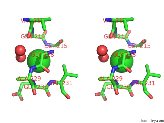
Stereo pair view
Mono view
Stereo pair view
A full contact list of Chlorine with other atoms in the Cl binding
site number 1 of Crystal Structure of Protein Phosphatase-1 Bound to the Natural Toxin Inhibitor Tautomycin within 5.0Å range:
|
Reference:
M.S.Kelker,
R.Page,
W.Peti.
Crystal Structures of Protein Phosphatase-1 Bound to Nodularin-R and Tautomycin: A Novel Scaffold For Structure-Based Drug Design of Serine/Threonine Phosphatase Inhibitors J.Mol.Biol. V. 385 11 2009.
ISSN: ISSN 0022-2836
PubMed: 18992256
DOI: 10.1016/J.JMB.2008.10.053
Page generated: Fri Jul 11 04:43:45 2025
ISSN: ISSN 0022-2836
PubMed: 18992256
DOI: 10.1016/J.JMB.2008.10.053
Last articles
Na in 3L6INa in 3L27
Na in 3L1O
Na in 3L0O
Na in 3KWM
Na in 3KYJ
Na in 3KQE
Na in 3KRS
Na in 3KW8
Na in 3KNT