Chlorine »
PDB 3fei-3fpl »
3flb »
Chlorine in PDB 3flb: Crystal Structure of Had Family Hydrolase DR_1622 From Deinococcus Radiodurans R1 (Target Efi-501256) with Bound Magnesium
Protein crystallography data
The structure of Crystal Structure of Had Family Hydrolase DR_1622 From Deinococcus Radiodurans R1 (Target Efi-501256) with Bound Magnesium, PDB code: 3flb
was solved by
Y.Patskovsky,
R.Toro,
R.Bhosle,
B.Hillerich,
R.D.Seidel,
E.Washington,
A.Scott Glenn,
S.Chowdhury,
B.Evans,
J.Hammonds,
W.D.Zencheck,
H.J.Imker,
J.A.Gerlt,
K.N.Allen,
D.Dunaway-Mariano,
S.C.Almo,
Enzyme Functioninitiative (Efi),
with X-Ray Crystallography technique. A brief refinement statistics is given in the table below:
| Resolution Low / High (Å) | 48.03 / 1.65 |
| Space group | P 65 2 2 |
| Cell size a, b, c (Å), α, β, γ (°) | 55.456, 55.456, 274.172, 90.00, 90.00, 120.00 |
| R / Rfree (%) | 20.2 / 23.5 |
Chlorine Binding Sites:
The binding sites of Chlorine atom in the Crystal Structure of Had Family Hydrolase DR_1622 From Deinococcus Radiodurans R1 (Target Efi-501256) with Bound Magnesium
(pdb code 3flb). This binding sites where shown within
5.0 Angstroms radius around Chlorine atom.
In total 3 binding sites of Chlorine where determined in the Crystal Structure of Had Family Hydrolase DR_1622 From Deinococcus Radiodurans R1 (Target Efi-501256) with Bound Magnesium, PDB code: 3flb:
Jump to Chlorine binding site number: 1; 2; 3;
In total 3 binding sites of Chlorine where determined in the Crystal Structure of Had Family Hydrolase DR_1622 From Deinococcus Radiodurans R1 (Target Efi-501256) with Bound Magnesium, PDB code: 3flb:
Jump to Chlorine binding site number: 1; 2; 3;
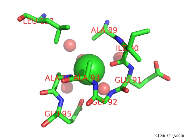
Chlorine binding site 1 out of 3 in 3flb
Go back to
Chlorine binding site 1 out
of 3 in the Crystal Structure of Had Family Hydrolase DR_1622 From Deinococcus Radiodurans R1 (Target Efi-501256) with Bound Magnesium
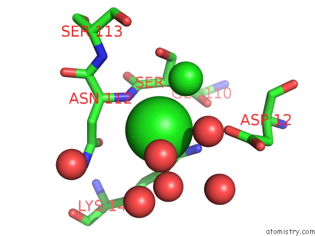
Mono view
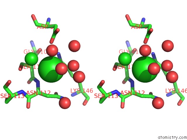
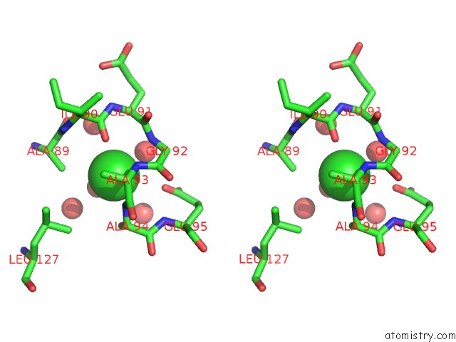
Stereo pair view
Mono view
Stereo pair view
A full contact list of Chlorine with other atoms in the Cl binding
site number 1 of Crystal Structure of Had Family Hydrolase DR_1622 From Deinococcus Radiodurans R1 (Target Efi-501256) with Bound Magnesium within 5.0Å range:
|
Chlorine binding site 2 out of 3 in 3flb
Go back to
Chlorine binding site 2 out
of 3 in the Crystal Structure of Had Family Hydrolase DR_1622 From Deinococcus Radiodurans R1 (Target Efi-501256) with Bound Magnesium
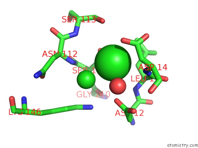
Mono view
Stereo pair view
Mono view
Stereo pair view
A full contact list of Chlorine with other atoms in the Cl binding
site number 2 of Crystal Structure of Had Family Hydrolase DR_1622 From Deinococcus Radiodurans R1 (Target Efi-501256) with Bound Magnesium within 5.0Å range:
|
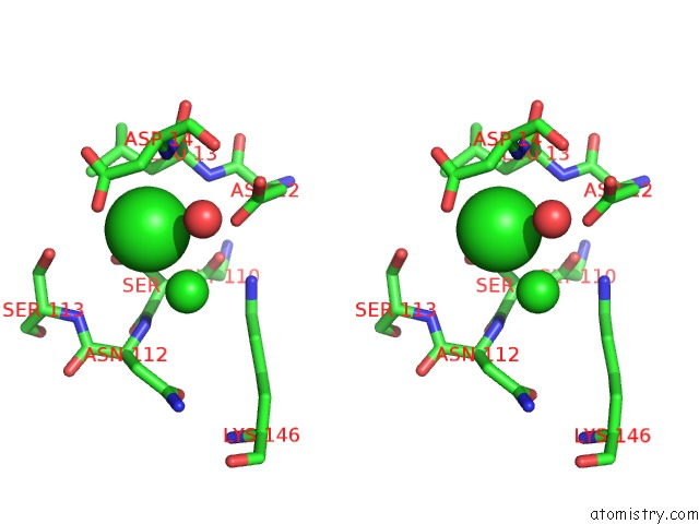
Chlorine binding site 3 out of 3 in 3flb
Go back to
Chlorine binding site 3 out
of 3 in the Crystal Structure of Had Family Hydrolase DR_1622 From Deinococcus Radiodurans R1 (Target Efi-501256) with Bound Magnesium
Mono view
Stereo pair view
Mono view
Stereo pair view
A full contact list of Chlorine with other atoms in the Cl binding
site number 3 of Crystal Structure of Had Family Hydrolase DR_1622 From Deinococcus Radiodurans R1 (Target Efi-501256) with Bound Magnesium within 5.0Å range:
|
Reference:
Y.Patskovsky,
R.Toro,
R.Bhosle,
B.Hillerich,
R.D.Seidel,
E.Washington,
A.Scott Glenn,
S.Chowdhury,
B.Evans,
J.Hammonds,
W.D.Zencheck,
H.J.Imker,
K.N.Allen,
D.Dunaway-Mariano,
J.A.Gerlt,
S.C.Almo.
Crystal Structure of Had Hydrolase DR_1622 Deinococcus Radiodurans R1 (Target Efi-501256) To Be Published.
Page generated: Fri Jul 11 05:08:49 2025
Last articles
Mn in 9LJUMn in 9LJW
Mn in 9LJS
Mn in 9LJR
Mn in 9LJT
Mn in 9LJV
Mg in 9UA2
Mg in 9R96
Mg in 9VM1
Mg in 9P01