Chlorine »
PDB 3fr1-3fzn »
3fwk »
Chlorine in PDB 3fwk: Crystal Structure of Candida Glabrata Fmn Adenylyltransferase
Enzymatic activity of Crystal Structure of Candida Glabrata Fmn Adenylyltransferase
All present enzymatic activity of Crystal Structure of Candida Glabrata Fmn Adenylyltransferase:
2.7.7.2;
2.7.7.2;
Protein crystallography data
The structure of Crystal Structure of Candida Glabrata Fmn Adenylyltransferase, PDB code: 3fwk
was solved by
C.Huerta,
D.Borek,
H.Zhang,
with X-Ray Crystallography technique. A brief refinement statistics is given in the table below:
| Resolution Low / High (Å) | 50.00 / 1.20 |
| Space group | P 32 2 1 |
| Cell size a, b, c (Å), α, β, γ (°) | 80.090, 80.090, 78.089, 90.00, 90.00, 120.00 |
| R / Rfree (%) | 16.2 / 17.8 |
Chlorine Binding Sites:
The binding sites of Chlorine atom in the Crystal Structure of Candida Glabrata Fmn Adenylyltransferase
(pdb code 3fwk). This binding sites where shown within
5.0 Angstroms radius around Chlorine atom.
In total only one binding site of Chlorine was determined in the Crystal Structure of Candida Glabrata Fmn Adenylyltransferase, PDB code: 3fwk:
In total only one binding site of Chlorine was determined in the Crystal Structure of Candida Glabrata Fmn Adenylyltransferase, PDB code: 3fwk:
Chlorine binding site 1 out of 1 in 3fwk
Go back to
Chlorine binding site 1 out
of 1 in the Crystal Structure of Candida Glabrata Fmn Adenylyltransferase
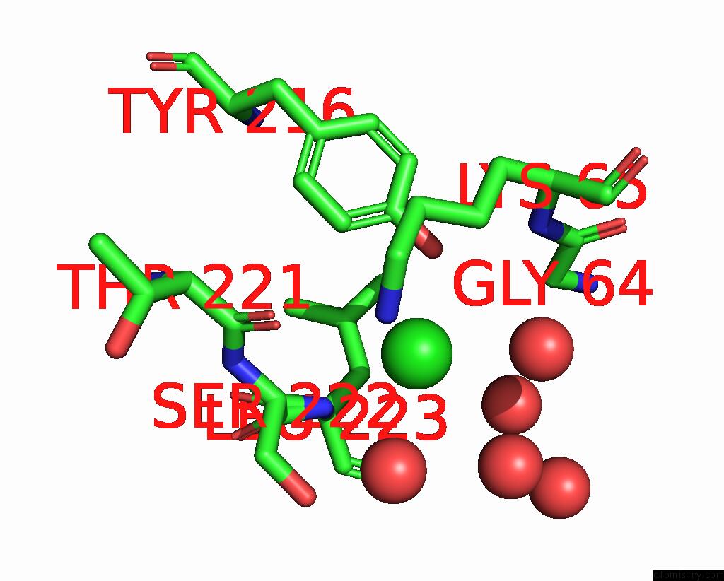
Mono view
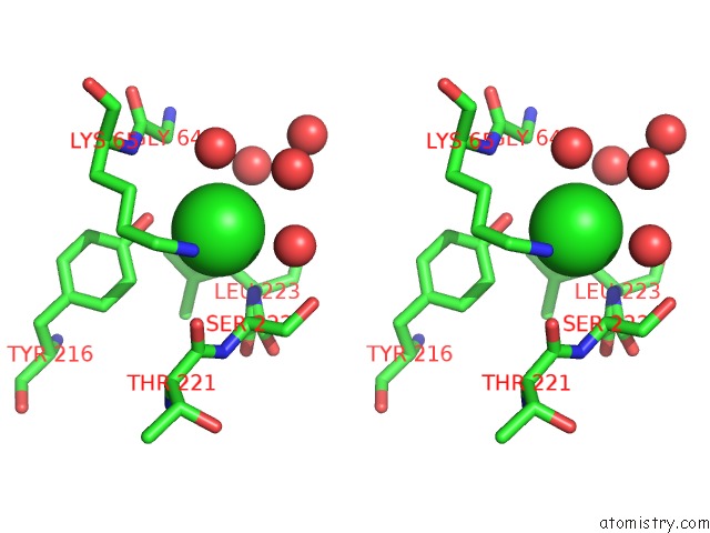
Stereo pair view
Mono view
Stereo pair view
A full contact list of Chlorine with other atoms in the Cl binding
site number 1 of Crystal Structure of Candida Glabrata Fmn Adenylyltransferase within 5.0Å range:
|
Reference:
C.Huerta,
D.Borek,
M.Machius,
N.V.Grishin,
H.Zhang.
Structure and Mechanism of A Eukaryotic Fmn Adenylyltransferase. J.Mol.Biol. V. 389 388 2009.
ISSN: ISSN 0022-2836
PubMed: 19375431
DOI: 10.1016/J.JMB.2009.04.022
Page generated: Fri Jul 11 05:16:34 2025
ISSN: ISSN 0022-2836
PubMed: 19375431
DOI: 10.1016/J.JMB.2009.04.022
Last articles
Mg in 6MW7Mg in 6MWK
Mg in 6MTI
Mg in 6MVE
Mg in 6MV9
Mg in 6MU4
Mg in 6MTH
Mg in 6MTA
Mg in 6MT8
Mg in 6MT9