Chlorine »
PDB 3g7q-3gjx »
3gba »
Chlorine in PDB 3gba: X-Ray Structure of IGLUR5 Ligand-Binding Core (S1S2) in Complex with Dysiherbaine at 1.35A Resolution
Protein crystallography data
The structure of X-Ray Structure of IGLUR5 Ligand-Binding Core (S1S2) in Complex with Dysiherbaine at 1.35A Resolution, PDB code: 3gba
was solved by
K.Frydenvang,
P.Naur,
M.Gajhede,
J.S.Kastrup,
with X-Ray Crystallography technique. A brief refinement statistics is given in the table below:
| Resolution Low / High (Å) | 89.80 / 1.35 |
| Space group | P 1 |
| Cell size a, b, c (Å), α, β, γ (°) | 44.872, 66.902, 90.342, 92.69, 94.66, 100.82 |
| R / Rfree (%) | 16.5 / 19.3 |
Chlorine Binding Sites:
The binding sites of Chlorine atom in the X-Ray Structure of IGLUR5 Ligand-Binding Core (S1S2) in Complex with Dysiherbaine at 1.35A Resolution
(pdb code 3gba). This binding sites where shown within
5.0 Angstroms radius around Chlorine atom.
In total 2 binding sites of Chlorine where determined in the X-Ray Structure of IGLUR5 Ligand-Binding Core (S1S2) in Complex with Dysiherbaine at 1.35A Resolution, PDB code: 3gba:
Jump to Chlorine binding site number: 1; 2;
In total 2 binding sites of Chlorine where determined in the X-Ray Structure of IGLUR5 Ligand-Binding Core (S1S2) in Complex with Dysiherbaine at 1.35A Resolution, PDB code: 3gba:
Jump to Chlorine binding site number: 1; 2;
Chlorine binding site 1 out of 2 in 3gba
Go back to
Chlorine binding site 1 out
of 2 in the X-Ray Structure of IGLUR5 Ligand-Binding Core (S1S2) in Complex with Dysiherbaine at 1.35A Resolution
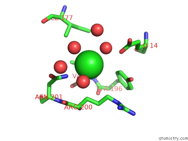
Mono view
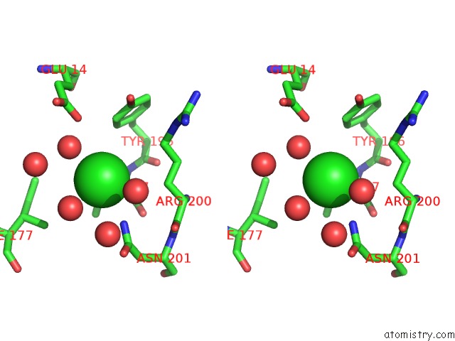
Stereo pair view
Mono view
Stereo pair view
A full contact list of Chlorine with other atoms in the Cl binding
site number 1 of X-Ray Structure of IGLUR5 Ligand-Binding Core (S1S2) in Complex with Dysiherbaine at 1.35A Resolution within 5.0Å range:
|
Chlorine binding site 2 out of 2 in 3gba
Go back to
Chlorine binding site 2 out
of 2 in the X-Ray Structure of IGLUR5 Ligand-Binding Core (S1S2) in Complex with Dysiherbaine at 1.35A Resolution
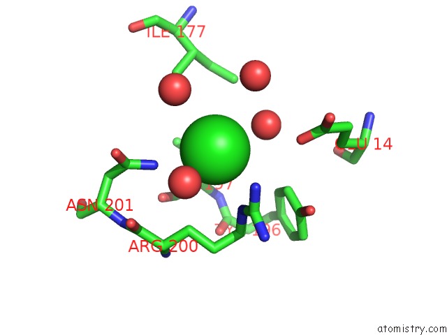
Mono view
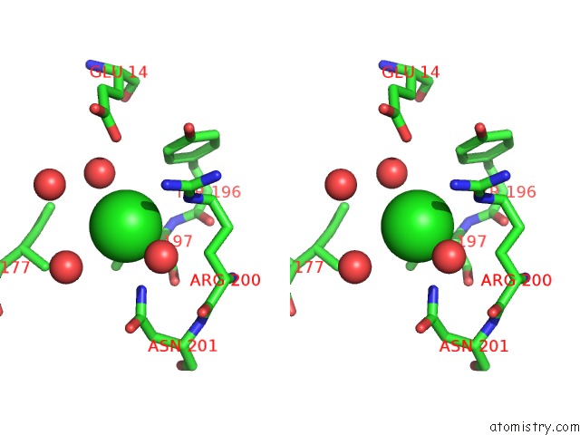
Stereo pair view
Mono view
Stereo pair view
A full contact list of Chlorine with other atoms in the Cl binding
site number 2 of X-Ray Structure of IGLUR5 Ligand-Binding Core (S1S2) in Complex with Dysiherbaine at 1.35A Resolution within 5.0Å range:
|
Reference:
K.Frydenvang,
L.L.Lash,
P.Naur,
P.A.Postila,
D.S.Pickering,
C.M.Smith,
M.Gajhede,
M.Sasaki,
R.Sakai,
O.T.Pentikainen,
G.T.Swanson,
J.S.Kastrup.
Full Domain Closure of the Ligand-Binding Core of the Ionotropic Glutamate Receptor IGLUR5 Induced By the High Affinity Agonist Dysiherbaine and the Functional Antagonist 8,9-Dideoxyneodysiherbaine J.Biol.Chem. V. 284 14219 2009.
ISSN: ISSN 0021-9258
PubMed: 19297335
DOI: 10.1074/JBC.M808547200
Page generated: Fri Jul 11 05:35:56 2025
ISSN: ISSN 0021-9258
PubMed: 19297335
DOI: 10.1074/JBC.M808547200
Last articles
Mg in 1ZZNMg in 207D
Mg in 1ZYK
Mg in 1ZXY
Mg in 1ZZ5
Mg in 1ZYR
Mg in 1ZYQ
Mg in 1ZYD
Mg in 1ZXN
Mg in 1ZY5