Chlorine »
PDB 3g7q-3gjx »
3geu »
Chlorine in PDB 3geu: Crystal Structure of Icar From Staphylococcus Aureus, A Member of the Tetracycline Repressor Protein Family
Protein crystallography data
The structure of Crystal Structure of Icar From Staphylococcus Aureus, A Member of the Tetracycline Repressor Protein Family, PDB code: 3geu
was solved by
S.M.Anderson,
J.S.Brunzelle,
Z.Wawrzak,
T.Skarina,
L.Papazisi,
W.F.Anderson,
A.Savchenko,
Center For Structural Genomics Ofinfectious Diseases (Csgid),
with X-Ray Crystallography technique. A brief refinement statistics is given in the table below:
| Resolution Low / High (Å) | 40.42 / 1.90 |
| Space group | P 21 21 2 |
| Cell size a, b, c (Å), α, β, γ (°) | 113.922, 121.141, 61.686, 90.00, 90.00, 90.00 |
| R / Rfree (%) | 18.2 / 22.8 |
Other elements in 3geu:
The structure of Crystal Structure of Icar From Staphylococcus Aureus, A Member of the Tetracycline Repressor Protein Family also contains other interesting chemical elements:
| Sodium | (Na) | 1 atom |
Chlorine Binding Sites:
The binding sites of Chlorine atom in the Crystal Structure of Icar From Staphylococcus Aureus, A Member of the Tetracycline Repressor Protein Family
(pdb code 3geu). This binding sites where shown within
5.0 Angstroms radius around Chlorine atom.
In total 6 binding sites of Chlorine where determined in the Crystal Structure of Icar From Staphylococcus Aureus, A Member of the Tetracycline Repressor Protein Family, PDB code: 3geu:
Jump to Chlorine binding site number: 1; 2; 3; 4; 5; 6;
In total 6 binding sites of Chlorine where determined in the Crystal Structure of Icar From Staphylococcus Aureus, A Member of the Tetracycline Repressor Protein Family, PDB code: 3geu:
Jump to Chlorine binding site number: 1; 2; 3; 4; 5; 6;
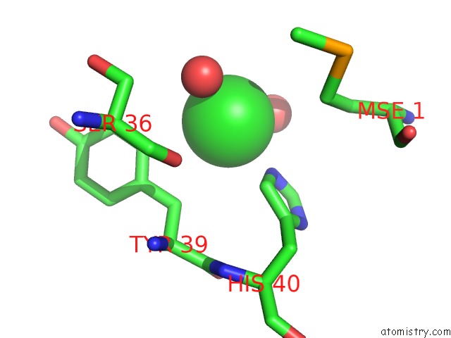
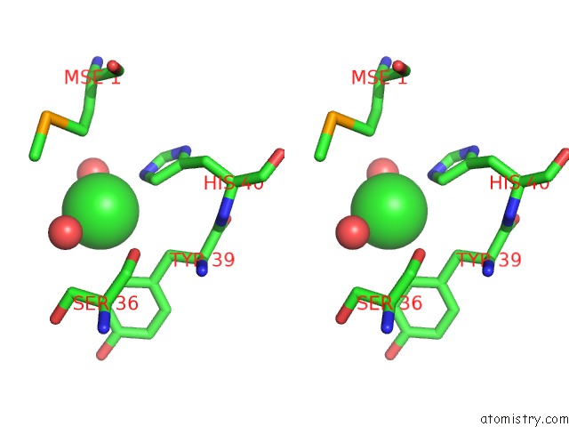
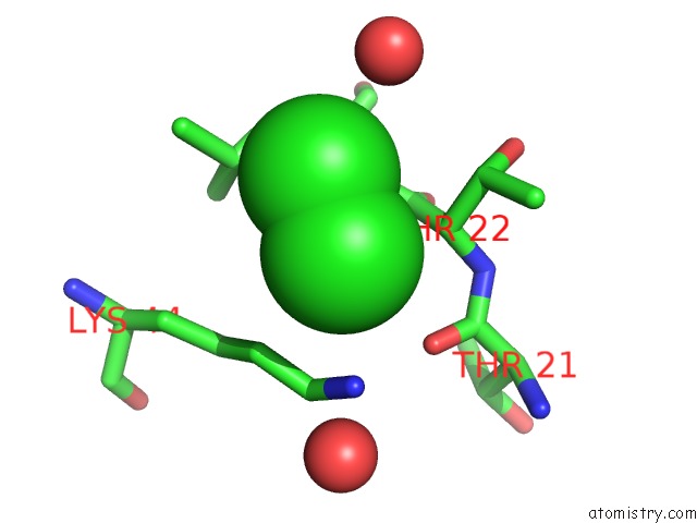
Chlorine binding site 1 out of 6 in 3geu
Go back to
Chlorine binding site 1 out
of 6 in the Crystal Structure of Icar From Staphylococcus Aureus, A Member of the Tetracycline Repressor Protein Family

Mono view

Stereo pair view
Mono view
Stereo pair view
A full contact list of Chlorine with other atoms in the Cl binding
site number 1 of Crystal Structure of Icar From Staphylococcus Aureus, A Member of the Tetracycline Repressor Protein Family within 5.0Å range:
|
Chlorine binding site 2 out of 6 in 3geu
Go back to
Chlorine binding site 2 out
of 6 in the Crystal Structure of Icar From Staphylococcus Aureus, A Member of the Tetracycline Repressor Protein Family
Mono view
Stereo pair view
Mono view
Stereo pair view
A full contact list of Chlorine with other atoms in the Cl binding
site number 2 of Crystal Structure of Icar From Staphylococcus Aureus, A Member of the Tetracycline Repressor Protein Family within 5.0Å range:
|
Chlorine binding site 3 out of 6 in 3geu
Go back to
Chlorine binding site 3 out
of 6 in the Crystal Structure of Icar From Staphylococcus Aureus, A Member of the Tetracycline Repressor Protein Family
Mono view
Stereo pair view
Mono view
Stereo pair view
A full contact list of Chlorine with other atoms in the Cl binding
site number 3 of Crystal Structure of Icar From Staphylococcus Aureus, A Member of the Tetracycline Repressor Protein Family within 5.0Å range:
|
Chlorine binding site 4 out of 6 in 3geu
Go back to
Chlorine binding site 4 out
of 6 in the Crystal Structure of Icar From Staphylococcus Aureus, A Member of the Tetracycline Repressor Protein Family

Mono view

Stereo pair view
Mono view
Stereo pair view
A full contact list of Chlorine with other atoms in the Cl binding
site number 4 of Crystal Structure of Icar From Staphylococcus Aureus, A Member of the Tetracycline Repressor Protein Family within 5.0Å range:
|
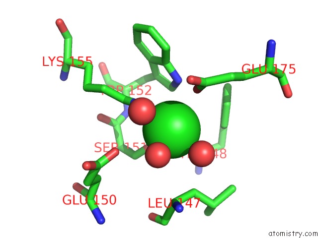
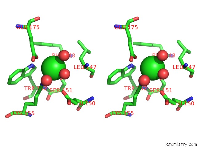
Chlorine binding site 5 out of 6 in 3geu
Go back to
Chlorine binding site 5 out
of 6 in the Crystal Structure of Icar From Staphylococcus Aureus, A Member of the Tetracycline Repressor Protein Family
Mono view

Stereo pair view
Mono view
Stereo pair view
A full contact list of Chlorine with other atoms in the Cl binding
site number 5 of Crystal Structure of Icar From Staphylococcus Aureus, A Member of the Tetracycline Repressor Protein Family within 5.0Å range:
|
Chlorine binding site 6 out of 6 in 3geu
Go back to
Chlorine binding site 6 out
of 6 in the Crystal Structure of Icar From Staphylococcus Aureus, A Member of the Tetracycline Repressor Protein Family

Mono view

Stereo pair view
Mono view
Stereo pair view
A full contact list of Chlorine with other atoms in the Cl binding
site number 6 of Crystal Structure of Icar From Staphylococcus Aureus, A Member of the Tetracycline Repressor Protein Family within 5.0Å range:
|
Reference:
S.M.Anderson,
J.S.Brunzelle,
Z.Wawrzak,
T.Skarina,
L.Papazisi,
W.F.Anderson,
A.Savchenko,
Center For Structural Genomics Of Infectious Diseases(Csgid).
Crystal Structure of Icar From Staphylococcus Aureus, A Member of the Tetracycline Repressor Protein Family To Be Published.
Page generated: Fri Jul 11 05:38:55 2025
Last articles
Mg in 4OX9Mg in 4P8R
Mg in 4P7A
Mg in 4P5J
Mg in 4P3E
Mg in 4P4S
Mg in 4P4O
Mg in 4P4M
Mg in 4P3Y
Mg in 4P3D