Chlorine »
PDB 3gky-3gxg »
3go5 »
Chlorine in PDB 3go5: Crystal Structure of A Multidomain Protein with Nucleic Acid Binding Domains (SP_0946) From Streptococcus Pneumoniae TIGR4 at 1.40 A Resolution
Protein crystallography data
The structure of Crystal Structure of A Multidomain Protein with Nucleic Acid Binding Domains (SP_0946) From Streptococcus Pneumoniae TIGR4 at 1.40 A Resolution, PDB code: 3go5
was solved by
Joint Center For Structural Genomics (Jcsg),
with X-Ray Crystallography technique. A brief refinement statistics is given in the table below:
| Resolution Low / High (Å) | 29.14 / 1.40 |
| Space group | P 32 2 1 |
| Cell size a, b, c (Å), α, β, γ (°) | 62.560, 62.560, 160.200, 90.00, 90.00, 120.00 |
| R / Rfree (%) | 15.4 / 17.5 |
Chlorine Binding Sites:
The binding sites of Chlorine atom in the Crystal Structure of A Multidomain Protein with Nucleic Acid Binding Domains (SP_0946) From Streptococcus Pneumoniae TIGR4 at 1.40 A Resolution
(pdb code 3go5). This binding sites where shown within
5.0 Angstroms radius around Chlorine atom.
In total only one binding site of Chlorine was determined in the Crystal Structure of A Multidomain Protein with Nucleic Acid Binding Domains (SP_0946) From Streptococcus Pneumoniae TIGR4 at 1.40 A Resolution, PDB code: 3go5:
In total only one binding site of Chlorine was determined in the Crystal Structure of A Multidomain Protein with Nucleic Acid Binding Domains (SP_0946) From Streptococcus Pneumoniae TIGR4 at 1.40 A Resolution, PDB code: 3go5:
Chlorine binding site 1 out of 1 in 3go5
Go back to
Chlorine binding site 1 out
of 1 in the Crystal Structure of A Multidomain Protein with Nucleic Acid Binding Domains (SP_0946) From Streptococcus Pneumoniae TIGR4 at 1.40 A Resolution
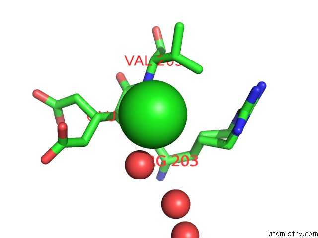
Mono view
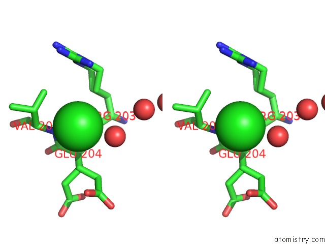
Stereo pair view
Mono view
Stereo pair view
A full contact list of Chlorine with other atoms in the Cl binding
site number 1 of Crystal Structure of A Multidomain Protein with Nucleic Acid Binding Domains (SP_0946) From Streptococcus Pneumoniae TIGR4 at 1.40 A Resolution within 5.0Å range:
|
Reference:
Y.Matsumoto,
Q.Xu,
S.Miyazaki,
C.Kaito,
C.L.Farr,
H.L.Axelrod,
H.J.Chiu,
H.E.Klock,
M.W.Knuth,
M.D.Miller,
M.A.Elsliger,
A.M.Deacon,
A.Godzik,
S.A.Lesley,
K.Sekimizu,
I.A.Wilson.
Structure of A Virulence Regulatory Factor Cvfb Reveals A Novel Winged Helix Rna Binding Module. Structure V. 18 537 2010.
ISSN: ISSN 0969-2126
PubMed: 20399190
DOI: 10.1016/J.STR.2010.02.007
Page generated: Fri Jul 11 05:43:41 2025
ISSN: ISSN 0969-2126
PubMed: 20399190
DOI: 10.1016/J.STR.2010.02.007
Last articles
Na in 6HZ0Na in 6HYZ
Na in 6HYX
Na in 6HYW
Na in 6HYV
Na in 6HY5
Na in 6HWS
Na in 6HWR
Na in 6HY8
Na in 6HXI