Chlorine »
PDB 3gky-3gxg »
3gug »
Chlorine in PDB 3gug: Crystal Structure of AKR1C1 L308V Mutant in Complex with Nadp and 3,5-Dichlorosalicylic Acid
Protein crystallography data
The structure of Crystal Structure of AKR1C1 L308V Mutant in Complex with Nadp and 3,5-Dichlorosalicylic Acid, PDB code: 3gug
was solved by
U.Dhagat,
O.El-Kabbani,
with X-Ray Crystallography technique. A brief refinement statistics is given in the table below:
| Resolution Low / High (Å) | 29.03 / 1.90 |
| Space group | P 1 21 1 |
| Cell size a, b, c (Å), α, β, γ (°) | 39.412, 83.885, 48.914, 90.00, 90.98, 90.00 |
| R / Rfree (%) | 19 / 26 |
Other elements in 3gug:
The structure of Crystal Structure of AKR1C1 L308V Mutant in Complex with Nadp and 3,5-Dichlorosalicylic Acid also contains other interesting chemical elements:
| Zinc | (Zn) | 1 atom |
Chlorine Binding Sites:
The binding sites of Chlorine atom in the Crystal Structure of AKR1C1 L308V Mutant in Complex with Nadp and 3,5-Dichlorosalicylic Acid
(pdb code 3gug). This binding sites where shown within
5.0 Angstroms radius around Chlorine atom.
In total 2 binding sites of Chlorine where determined in the Crystal Structure of AKR1C1 L308V Mutant in Complex with Nadp and 3,5-Dichlorosalicylic Acid, PDB code: 3gug:
Jump to Chlorine binding site number: 1; 2;
In total 2 binding sites of Chlorine where determined in the Crystal Structure of AKR1C1 L308V Mutant in Complex with Nadp and 3,5-Dichlorosalicylic Acid, PDB code: 3gug:
Jump to Chlorine binding site number: 1; 2;
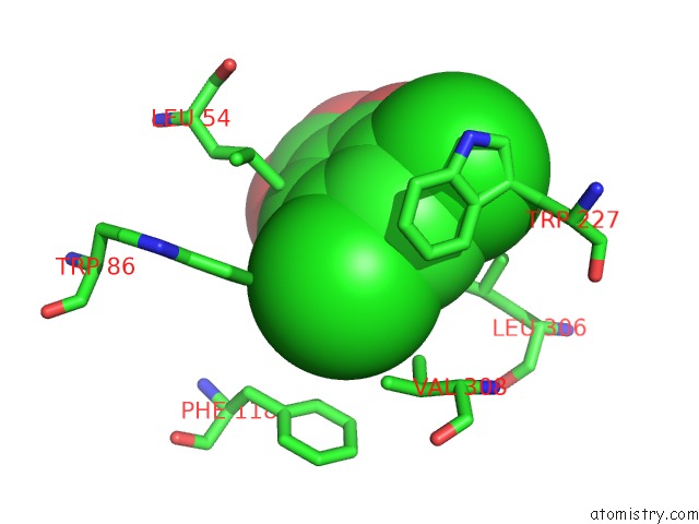
Chlorine binding site 1 out of 2 in 3gug
Go back to
Chlorine binding site 1 out
of 2 in the Crystal Structure of AKR1C1 L308V Mutant in Complex with Nadp and 3,5-Dichlorosalicylic Acid

Mono view

Stereo pair view
Mono view
Stereo pair view
A full contact list of Chlorine with other atoms in the Cl binding
site number 1 of Crystal Structure of AKR1C1 L308V Mutant in Complex with Nadp and 3,5-Dichlorosalicylic Acid within 5.0Å range:
|
Chlorine binding site 2 out of 2 in 3gug
Go back to
Chlorine binding site 2 out
of 2 in the Crystal Structure of AKR1C1 L308V Mutant in Complex with Nadp and 3,5-Dichlorosalicylic Acid
Mono view

Stereo pair view
Mono view
Stereo pair view
A full contact list of Chlorine with other atoms in the Cl binding
site number 2 of Crystal Structure of AKR1C1 L308V Mutant in Complex with Nadp and 3,5-Dichlorosalicylic Acid within 5.0Å range:
|
Reference:
U.Dhagat,
A.Hara,
O.El-Kabbani.
Crystal Structure of AKR1C1 L308V Mutant in Complex with Nadp and 3,5-Dichlorosalicylic Acid To Be Published.
Page generated: Fri Jul 11 05:46:29 2025
Last articles
Zn in 4HKIZn in 4HK6
Zn in 4HJW
Zn in 4HJE
Zn in 4HIF
Zn in 4HI9
Zn in 4HI8
Zn in 4HEX
Zn in 4HHJ
Zn in 4HF4