Chlorine »
PDB 3gxh-3hcx »
3h4w »
Chlorine in PDB 3h4w: Structure of A Ca+2 Dependent Phosphatidylinositol-Specific Phospholipase C (Pi-Plc) Enzyme From Streptomyces Antibioticus
Enzymatic activity of Structure of A Ca+2 Dependent Phosphatidylinositol-Specific Phospholipase C (Pi-Plc) Enzyme From Streptomyces Antibioticus
All present enzymatic activity of Structure of A Ca+2 Dependent Phosphatidylinositol-Specific Phospholipase C (Pi-Plc) Enzyme From Streptomyces Antibioticus:
3.1.4.11;
3.1.4.11;
Protein crystallography data
The structure of Structure of A Ca+2 Dependent Phosphatidylinositol-Specific Phospholipase C (Pi-Plc) Enzyme From Streptomyces Antibioticus, PDB code: 3h4w
was solved by
M.R.Jackson,
T.L.Selby,
with X-Ray Crystallography technique. A brief refinement statistics is given in the table below:
| Resolution Low / High (Å) | 77.59 / 1.50 |
| Space group | P 21 21 2 |
| Cell size a, b, c (Å), α, β, γ (°) | 52.377, 155.172, 41.409, 90.00, 90.00, 90.00 |
| R / Rfree (%) | 18.4 / 20 |
Chlorine Binding Sites:
The binding sites of Chlorine atom in the Structure of A Ca+2 Dependent Phosphatidylinositol-Specific Phospholipase C (Pi-Plc) Enzyme From Streptomyces Antibioticus
(pdb code 3h4w). This binding sites where shown within
5.0 Angstroms radius around Chlorine atom.
In total only one binding site of Chlorine was determined in the Structure of A Ca+2 Dependent Phosphatidylinositol-Specific Phospholipase C (Pi-Plc) Enzyme From Streptomyces Antibioticus, PDB code: 3h4w:
In total only one binding site of Chlorine was determined in the Structure of A Ca+2 Dependent Phosphatidylinositol-Specific Phospholipase C (Pi-Plc) Enzyme From Streptomyces Antibioticus, PDB code: 3h4w:
Chlorine binding site 1 out of 1 in 3h4w
Go back to
Chlorine binding site 1 out
of 1 in the Structure of A Ca+2 Dependent Phosphatidylinositol-Specific Phospholipase C (Pi-Plc) Enzyme From Streptomyces Antibioticus
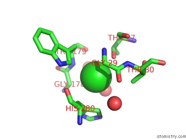
Mono view
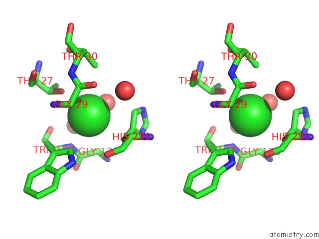
Stereo pair view
Mono view
Stereo pair view
A full contact list of Chlorine with other atoms in the Cl binding
site number 1 of Structure of A Ca+2 Dependent Phosphatidylinositol-Specific Phospholipase C (Pi-Plc) Enzyme From Streptomyces Antibioticus within 5.0Å range:
|
Reference:
M.R.Jackson,
T.L.Selby.
Crystal Structure of A CA2+-Dependent Pi-Plc To Be Published.
Page generated: Fri Jul 11 05:51:04 2025
Last articles
Mn in 2QULMn in 2QKC
Mn in 2QKA
Mn in 2QJC
Mn in 2QC8
Mn in 2QF2
Mn in 2QGI
Mn in 2QEY
Mn in 2QF1
Mn in 2QCS