Chlorine »
PDB 3gxh-3hcx »
3h8d »
Chlorine in PDB 3h8d: Crystal Structure of Myosin VI in Complex with DAB2 Peptide
Protein crystallography data
The structure of Crystal Structure of Myosin VI in Complex with DAB2 Peptide, PDB code: 3h8d
was solved by
C.Yu,
W.Feng,
Z.Wei,
M.Zhang,
with X-Ray Crystallography technique. A brief refinement statistics is given in the table below:
| Resolution Low / High (Å) | 30.00 / 2.20 |
| Space group | P 1 21 1 |
| Cell size a, b, c (Å), α, β, γ (°) | 69.177, 69.952, 78.537, 90.00, 98.02, 90.00 |
| R / Rfree (%) | 18.9 / 25.1 |
Chlorine Binding Sites:
The binding sites of Chlorine atom in the Crystal Structure of Myosin VI in Complex with DAB2 Peptide
(pdb code 3h8d). This binding sites where shown within
5.0 Angstroms radius around Chlorine atom.
In total only one binding site of Chlorine was determined in the Crystal Structure of Myosin VI in Complex with DAB2 Peptide, PDB code: 3h8d:
In total only one binding site of Chlorine was determined in the Crystal Structure of Myosin VI in Complex with DAB2 Peptide, PDB code: 3h8d:
Chlorine binding site 1 out of 1 in 3h8d
Go back to
Chlorine binding site 1 out
of 1 in the Crystal Structure of Myosin VI in Complex with DAB2 Peptide
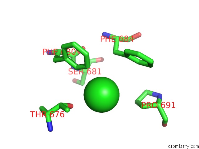
Mono view
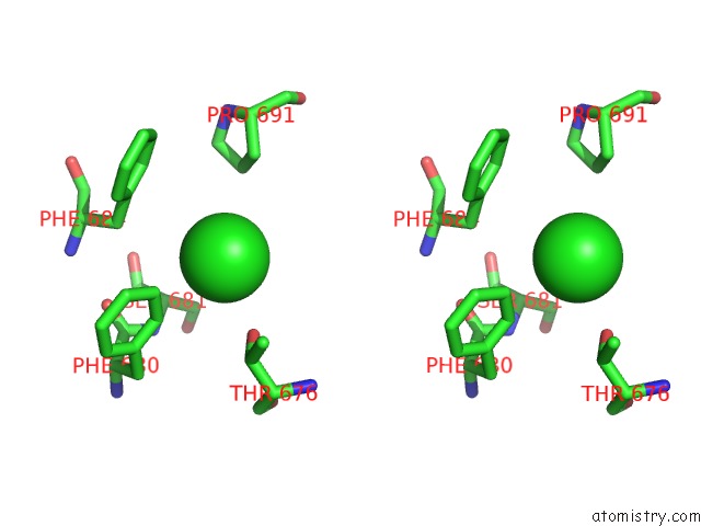
Stereo pair view
Mono view
Stereo pair view
A full contact list of Chlorine with other atoms in the Cl binding
site number 1 of Crystal Structure of Myosin VI in Complex with DAB2 Peptide within 5.0Å range:
|
Reference:
C.Yu,
W.Feng,
Z.Wei,
Y.Miyanoiri,
W.Wen,
Y.Zhao,
M.Zhang.
Myosin VI Undergoes Cargo-Mediated Dimerization Cell(Cambridge,Mass.) V. 138 537 2009.
ISSN: ISSN 0092-8674
PubMed: 19665975
DOI: 10.1016/J.CELL.2009.05.030
Page generated: Fri Jul 11 05:52:38 2025
ISSN: ISSN 0092-8674
PubMed: 19665975
DOI: 10.1016/J.CELL.2009.05.030
Last articles
Mn in 2QJTMn in 2R21
Mn in 2QUM
Mn in 2QUN
Mn in 2QUL
Mn in 2QKC
Mn in 2QKA
Mn in 2QJC
Mn in 2QC8
Mn in 2QF2