Chlorine »
PDB 3hd1-3hl7 »
3hhd »
Chlorine in PDB 3hhd: Structure of the Human Fatty Acid Synthase Ks-Mat Didomain As A Framework For Inhibitor Design.
Enzymatic activity of Structure of the Human Fatty Acid Synthase Ks-Mat Didomain As A Framework For Inhibitor Design.
All present enzymatic activity of Structure of the Human Fatty Acid Synthase Ks-Mat Didomain As A Framework For Inhibitor Design.:
2.3.1.85;
2.3.1.85;
Protein crystallography data
The structure of Structure of the Human Fatty Acid Synthase Ks-Mat Didomain As A Framework For Inhibitor Design., PDB code: 3hhd
was solved by
G.M.Pappenberger,
J.Benz,
R.Thoma,
M.G.Rudolph,
with X-Ray Crystallography technique. A brief refinement statistics is given in the table below:
| Resolution Low / High (Å) | 45.64 / 2.15 |
| Space group | P 1 |
| Cell size a, b, c (Å), α, β, γ (°) | 86.620, 91.160, 132.110, 73.84, 86.83, 62.54 |
| R / Rfree (%) | 16.9 / 21.1 |
Chlorine Binding Sites:
The binding sites of Chlorine atom in the Structure of the Human Fatty Acid Synthase Ks-Mat Didomain As A Framework For Inhibitor Design.
(pdb code 3hhd). This binding sites where shown within
5.0 Angstroms radius around Chlorine atom.
In total 2 binding sites of Chlorine where determined in the Structure of the Human Fatty Acid Synthase Ks-Mat Didomain As A Framework For Inhibitor Design., PDB code: 3hhd:
Jump to Chlorine binding site number: 1; 2;
In total 2 binding sites of Chlorine where determined in the Structure of the Human Fatty Acid Synthase Ks-Mat Didomain As A Framework For Inhibitor Design., PDB code: 3hhd:
Jump to Chlorine binding site number: 1; 2;
Chlorine binding site 1 out of 2 in 3hhd
Go back to
Chlorine binding site 1 out
of 2 in the Structure of the Human Fatty Acid Synthase Ks-Mat Didomain As A Framework For Inhibitor Design.
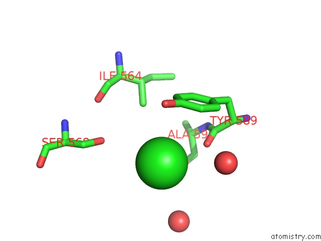
Mono view
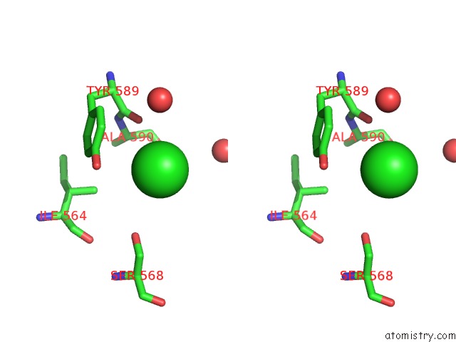
Stereo pair view
Mono view
Stereo pair view
A full contact list of Chlorine with other atoms in the Cl binding
site number 1 of Structure of the Human Fatty Acid Synthase Ks-Mat Didomain As A Framework For Inhibitor Design. within 5.0Å range:
|
Chlorine binding site 2 out of 2 in 3hhd
Go back to
Chlorine binding site 2 out
of 2 in the Structure of the Human Fatty Acid Synthase Ks-Mat Didomain As A Framework For Inhibitor Design.
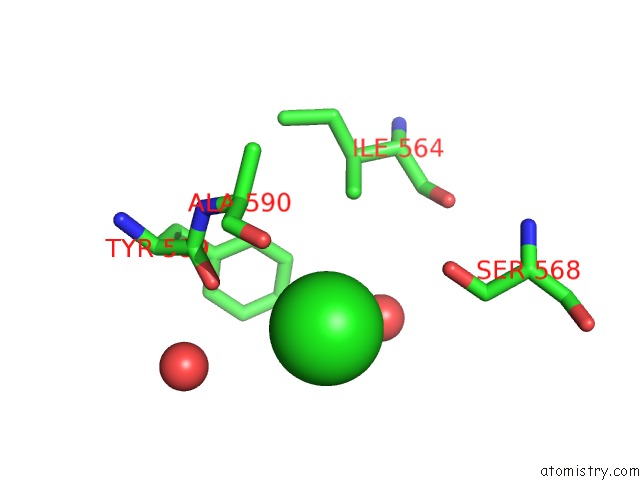
Mono view
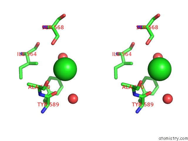
Stereo pair view
Mono view
Stereo pair view
A full contact list of Chlorine with other atoms in the Cl binding
site number 2 of Structure of the Human Fatty Acid Synthase Ks-Mat Didomain As A Framework For Inhibitor Design. within 5.0Å range:
|
Reference:
G.Pappenberger,
J.Benz,
B.Gsell,
M.Hennig,
A.Ruf,
M.Stihle,
R.Thoma,
M.G.Rudolph.
Structure of the Human Fatty Acid Synthase Ks-Mat Didomain As A Framework For Inhibitor Design. J.Mol.Biol. V. 397 508 2010.
ISSN: ISSN 0022-2836
PubMed: 20132826
DOI: 10.1016/J.JMB.2010.01.066
Page generated: Fri Jul 11 05:57:02 2025
ISSN: ISSN 0022-2836
PubMed: 20132826
DOI: 10.1016/J.JMB.2010.01.066
Last articles
Mn in 9LJUMn in 9LJW
Mn in 9LJS
Mn in 9LJR
Mn in 9LJT
Mn in 9LJV
Mg in 9UA2
Mg in 9R96
Mg in 9VM1
Mg in 9P01