Chlorine »
PDB 3hd1-3hl7 »
3hl7 »
Chlorine in PDB 3hl7: Crystal Structure of Human P38ALPHA Complexed with Sd-0006
Enzymatic activity of Crystal Structure of Human P38ALPHA Complexed with Sd-0006
All present enzymatic activity of Crystal Structure of Human P38ALPHA Complexed with Sd-0006:
2.7.11.24;
2.7.11.24;
Protein crystallography data
The structure of Crystal Structure of Human P38ALPHA Complexed with Sd-0006, PDB code: 3hl7
was solved by
H.-S.Shieh,
R.G.Kurumbail,
R.A.Stegeman,
J.M.Williams,
with X-Ray Crystallography technique. A brief refinement statistics is given in the table below:
| Resolution Low / High (Å) | 24.84 / 1.88 |
| Space group | P 21 21 21 |
| Cell size a, b, c (Å), α, β, γ (°) | 65.088, 74.525, 77.587, 90.00, 90.00, 90.00 |
| R / Rfree (%) | 19.9 / 22.8 |
Other elements in 3hl7:
The structure of Crystal Structure of Human P38ALPHA Complexed with Sd-0006 also contains other interesting chemical elements:
| Fluorine | (F) | 2 atoms |
Chlorine Binding Sites:
The binding sites of Chlorine atom in the Crystal Structure of Human P38ALPHA Complexed with Sd-0006
(pdb code 3hl7). This binding sites where shown within
5.0 Angstroms radius around Chlorine atom.
In total only one binding site of Chlorine was determined in the Crystal Structure of Human P38ALPHA Complexed with Sd-0006, PDB code: 3hl7:
In total only one binding site of Chlorine was determined in the Crystal Structure of Human P38ALPHA Complexed with Sd-0006, PDB code: 3hl7:
Chlorine binding site 1 out of 1 in 3hl7
Go back to
Chlorine binding site 1 out
of 1 in the Crystal Structure of Human P38ALPHA Complexed with Sd-0006
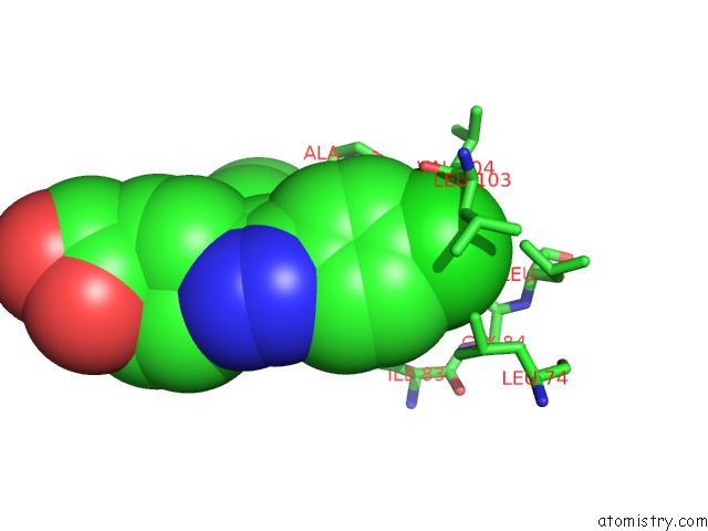
Mono view
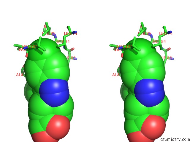
Stereo pair view
Mono view
Stereo pair view
A full contact list of Chlorine with other atoms in the Cl binding
site number 1 of Crystal Structure of Human P38ALPHA Complexed with Sd-0006 within 5.0Å range:
|
Reference:
L.Xing,
H.S.Shieh,
S.R.Selness,
R.V.Devraj,
J.K.Walker,
B.Devadas,
H.R.Hope,
R.P.Compton,
J.F.Schindler,
J.L.Hirsch,
A.G.Benson,
R.G.Kurumbail,
R.A.Stegeman,
J.M.Williams,
R.M.Broadus,
Z.Walden,
J.B.Monahan.
Structural Bioinformatics-Based Prediction of Exceptional Selectivity of P38 Map Kinase Inhibitor pH-797804. Biochemistry V. 48 6402 2009.
ISSN: ISSN 0006-2960
PubMed: 19496616
DOI: 10.1021/BI900655F
Page generated: Fri Jul 11 06:02:17 2025
ISSN: ISSN 0006-2960
PubMed: 19496616
DOI: 10.1021/BI900655F
Last articles
Mn in 9LJUMn in 9LJW
Mn in 9LJS
Mn in 9LJR
Mn in 9LJT
Mn in 9LJV
Mg in 9UA2
Mg in 9R96
Mg in 9VM1
Mg in 9P01