Chlorine »
PDB 3hl8-3hwl »
3hsz »
Chlorine in PDB 3hsz: Crystal Structure of E. Coli Hppk(F123A)
Enzymatic activity of Crystal Structure of E. Coli Hppk(F123A)
All present enzymatic activity of Crystal Structure of E. Coli Hppk(F123A):
2.7.6.3;
2.7.6.3;
Protein crystallography data
The structure of Crystal Structure of E. Coli Hppk(F123A), PDB code: 3hsz
was solved by
J.Blaszczyk,
Y.Li,
H.Yan,
X.Ji,
with X-Ray Crystallography technique. A brief refinement statistics is given in the table below:
| Resolution Low / High (Å) | 21.96 / 1.40 |
| Space group | P 1 21 1 |
| Cell size a, b, c (Å), α, β, γ (°) | 36.150, 52.060, 41.390, 90.00, 110.54, 90.00 |
| R / Rfree (%) | 15 / 18.9 |
Chlorine Binding Sites:
The binding sites of Chlorine atom in the Crystal Structure of E. Coli Hppk(F123A)
(pdb code 3hsz). This binding sites where shown within
5.0 Angstroms radius around Chlorine atom.
In total only one binding site of Chlorine was determined in the Crystal Structure of E. Coli Hppk(F123A), PDB code: 3hsz:
In total only one binding site of Chlorine was determined in the Crystal Structure of E. Coli Hppk(F123A), PDB code: 3hsz:
Chlorine binding site 1 out of 1 in 3hsz
Go back to
Chlorine binding site 1 out
of 1 in the Crystal Structure of E. Coli Hppk(F123A)
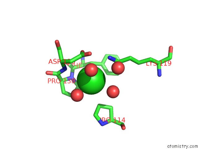
Mono view
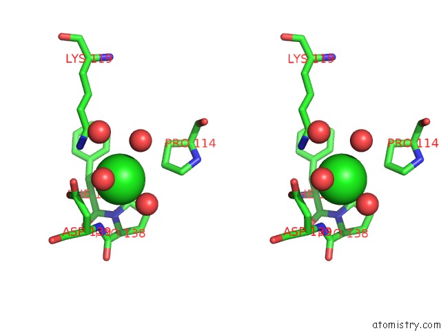
Stereo pair view
Mono view
Stereo pair view
A full contact list of Chlorine with other atoms in the Cl binding
site number 1 of Crystal Structure of E. Coli Hppk(F123A) within 5.0Å range:
|
Reference:
Y.Li,
J.Blaszczyk,
X.Ji,
H.Yan.
Pterin-Binding Site Mutation Y53A, N55A or F123A and Activity of E. Coli Hppk To Be Published.
Page generated: Fri Jul 11 06:04:58 2025
Last articles
Na in 1VI6Na in 1VKG
Na in 1VMJ
Na in 1VMH
Na in 1VMF
Na in 1VLM
Na in 1VK1
Na in 1VIZ
Na in 1VEL
Na in 1VE8