Chlorine »
PDB 3hwp-3i67 »
3i2a »
Chlorine in PDB 3i2a: Crystal Structure of A Chimeric Trypsin Inhibitor Protein Sti(L)- Wci(S)
Protein crystallography data
The structure of Crystal Structure of A Chimeric Trypsin Inhibitor Protein Sti(L)- Wci(S), PDB code: 3i2a
was solved by
U.Sen,
S.Khamrui,
with X-Ray Crystallography technique. A brief refinement statistics is given in the table below:
| Resolution Low / High (Å) | 19.96 / 2.30 |
| Space group | I 4 |
| Cell size a, b, c (Å), α, β, γ (°) | 141.383, 141.383, 46.716, 90.00, 90.00, 90.00 |
| R / Rfree (%) | 23.4 / 28.9 |
Chlorine Binding Sites:
The binding sites of Chlorine atom in the Crystal Structure of A Chimeric Trypsin Inhibitor Protein Sti(L)- Wci(S)
(pdb code 3i2a). This binding sites where shown within
5.0 Angstroms radius around Chlorine atom.
In total 2 binding sites of Chlorine where determined in the Crystal Structure of A Chimeric Trypsin Inhibitor Protein Sti(L)- Wci(S), PDB code: 3i2a:
Jump to Chlorine binding site number: 1; 2;
In total 2 binding sites of Chlorine where determined in the Crystal Structure of A Chimeric Trypsin Inhibitor Protein Sti(L)- Wci(S), PDB code: 3i2a:
Jump to Chlorine binding site number: 1; 2;
Chlorine binding site 1 out of 2 in 3i2a
Go back to
Chlorine binding site 1 out
of 2 in the Crystal Structure of A Chimeric Trypsin Inhibitor Protein Sti(L)- Wci(S)
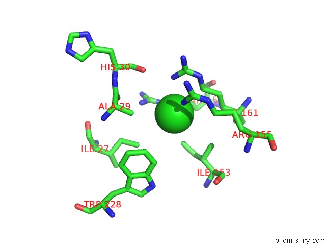
Mono view
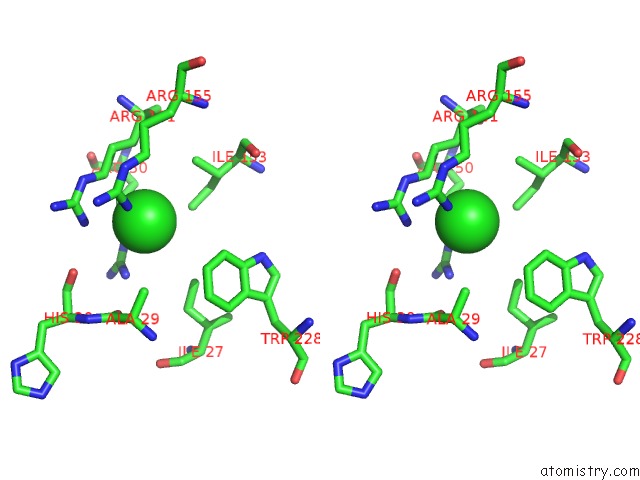
Stereo pair view
Mono view
Stereo pair view
A full contact list of Chlorine with other atoms in the Cl binding
site number 1 of Crystal Structure of A Chimeric Trypsin Inhibitor Protein Sti(L)- Wci(S) within 5.0Å range:
|
Chlorine binding site 2 out of 2 in 3i2a
Go back to
Chlorine binding site 2 out
of 2 in the Crystal Structure of A Chimeric Trypsin Inhibitor Protein Sti(L)- Wci(S)
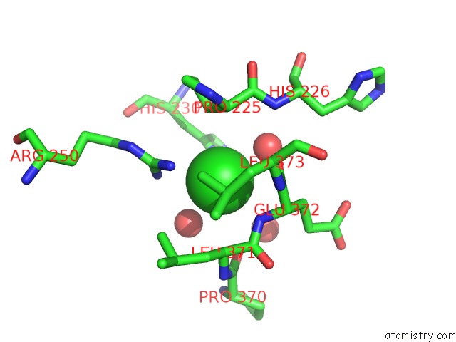
Mono view
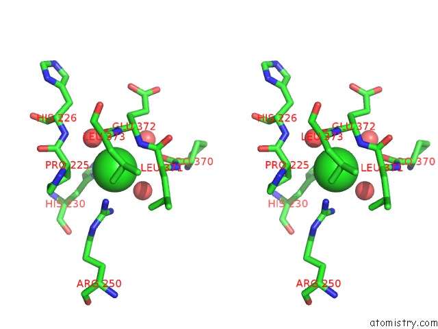
Stereo pair view
Mono view
Stereo pair view
A full contact list of Chlorine with other atoms in the Cl binding
site number 2 of Crystal Structure of A Chimeric Trypsin Inhibitor Protein Sti(L)- Wci(S) within 5.0Å range:
|
Reference:
S.Khamrui,
S.Majumder,
J.Dasgupta,
J.K.Dattagupta,
U.Sen.
Identification of A Novel Set of Scaffolding Residues That Are Instrumental For the Inhibitory Property of Kunitz (Sti) Inhibitors. Protein Sci. V. 19 593 2010.
ISSN: ISSN 0961-8368
PubMed: 20073082
DOI: 10.1002/PRO.338
Page generated: Fri Jul 11 06:09:41 2025
ISSN: ISSN 0961-8368
PubMed: 20073082
DOI: 10.1002/PRO.338
Last articles
F in 4GVHF in 4GU9
F in 4GSI
F in 4GPT
F in 4GTO
F in 4GS9
F in 4GQ4
F in 4GPB
F in 4GS0
F in 4GLU