Chlorine »
PDB 3ilj-3isv »
3ilj »
Chlorine in PDB 3ilj: Crystal Structure of E. Coli Hppk(D95A) in Complex with Mgampcpp
Enzymatic activity of Crystal Structure of E. Coli Hppk(D95A) in Complex with Mgampcpp
All present enzymatic activity of Crystal Structure of E. Coli Hppk(D95A) in Complex with Mgampcpp:
2.7.6.3;
2.7.6.3;
Protein crystallography data
The structure of Crystal Structure of E. Coli Hppk(D95A) in Complex with Mgampcpp, PDB code: 3ilj
was solved by
J.Blaszczyk,
Y.Li,
H.Yan,
X.Ji,
with X-Ray Crystallography technique. A brief refinement statistics is given in the table below:
| Resolution Low / High (Å) | 29.27 / 1.65 |
| Space group | P 21 21 2 |
| Cell size a, b, c (Å), α, β, γ (°) | 53.030, 70.210, 36.260, 90.00, 90.00, 90.00 |
| R / Rfree (%) | 18.2 / 22.5 |
Other elements in 3ilj:
The structure of Crystal Structure of E. Coli Hppk(D95A) in Complex with Mgampcpp also contains other interesting chemical elements:
| Magnesium | (Mg) | 1 atom |
Chlorine Binding Sites:
The binding sites of Chlorine atom in the Crystal Structure of E. Coli Hppk(D95A) in Complex with Mgampcpp
(pdb code 3ilj). This binding sites where shown within
5.0 Angstroms radius around Chlorine atom.
In total only one binding site of Chlorine was determined in the Crystal Structure of E. Coli Hppk(D95A) in Complex with Mgampcpp, PDB code: 3ilj:
In total only one binding site of Chlorine was determined in the Crystal Structure of E. Coli Hppk(D95A) in Complex with Mgampcpp, PDB code: 3ilj:
Chlorine binding site 1 out of 1 in 3ilj
Go back to
Chlorine binding site 1 out
of 1 in the Crystal Structure of E. Coli Hppk(D95A) in Complex with Mgampcpp
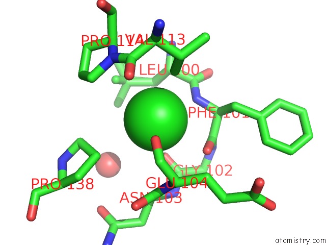
Mono view
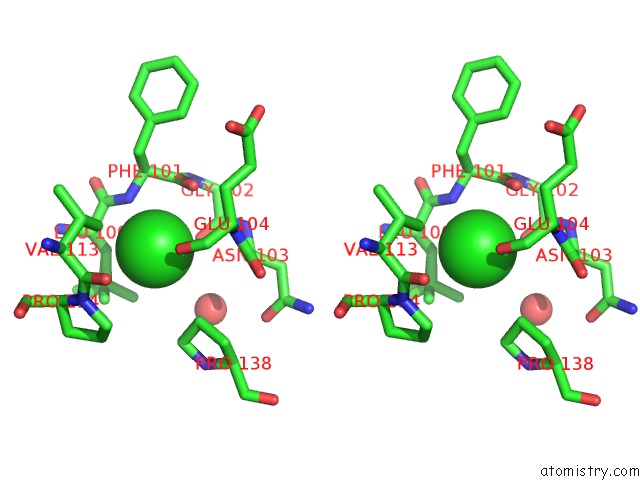
Stereo pair view
Mono view
Stereo pair view
A full contact list of Chlorine with other atoms in the Cl binding
site number 1 of Crystal Structure of E. Coli Hppk(D95A) in Complex with Mgampcpp within 5.0Å range:
|
Reference:
Y.Li,
J.Blaszczyk,
X.Ji,
H.Yan.
Structural and Functional Roles of Residues D95 and D97 in E. Coli Hppk To Be Published.
Page generated: Fri Jul 11 06:26:04 2025
Last articles
Mg in 6QV0Mg in 6QUZ
Mg in 6QUY
Mg in 6QUX
Mg in 6QUW
Mg in 6QUV
Mg in 6QUU
Mg in 6QUS
Mg in 6QTN
Mg in 6QUM