Chlorine »
PDB 3itd-3jpy »
3ivr »
Chlorine in PDB 3ivr: Crystal Structure of Putative Long-Chain-Fatty-Acid Coa Ligase From Rhodopseudomonas Palustris CGA009
Enzymatic activity of Crystal Structure of Putative Long-Chain-Fatty-Acid Coa Ligase From Rhodopseudomonas Palustris CGA009
All present enzymatic activity of Crystal Structure of Putative Long-Chain-Fatty-Acid Coa Ligase From Rhodopseudomonas Palustris CGA009:
6.2.1.3;
6.2.1.3;
Protein crystallography data
The structure of Crystal Structure of Putative Long-Chain-Fatty-Acid Coa Ligase From Rhodopseudomonas Palustris CGA009, PDB code: 3ivr
was solved by
Y.Patskovsky,
R.Toro,
R.Foti,
M.Dickey,
J.M.Sauder,
S.K.Burley,
S.C.Almo,
New York Sgx Research Center For Structural Genomics (Nysgxrc),
with X-Ray Crystallography technique. A brief refinement statistics is given in the table below:
| Resolution Low / High (Å) | 20.00 / 2.00 |
| Space group | C 1 2 1 |
| Cell size a, b, c (Å), α, β, γ (°) | 127.624, 97.213, 95.077, 90.00, 102.04, 90.00 |
| R / Rfree (%) | 17.3 / 21.7 |
Chlorine Binding Sites:
The binding sites of Chlorine atom in the Crystal Structure of Putative Long-Chain-Fatty-Acid Coa Ligase From Rhodopseudomonas Palustris CGA009
(pdb code 3ivr). This binding sites where shown within
5.0 Angstroms radius around Chlorine atom.
In total only one binding site of Chlorine was determined in the Crystal Structure of Putative Long-Chain-Fatty-Acid Coa Ligase From Rhodopseudomonas Palustris CGA009, PDB code: 3ivr:
In total only one binding site of Chlorine was determined in the Crystal Structure of Putative Long-Chain-Fatty-Acid Coa Ligase From Rhodopseudomonas Palustris CGA009, PDB code: 3ivr:
Chlorine binding site 1 out of 1 in 3ivr
Go back to
Chlorine binding site 1 out
of 1 in the Crystal Structure of Putative Long-Chain-Fatty-Acid Coa Ligase From Rhodopseudomonas Palustris CGA009
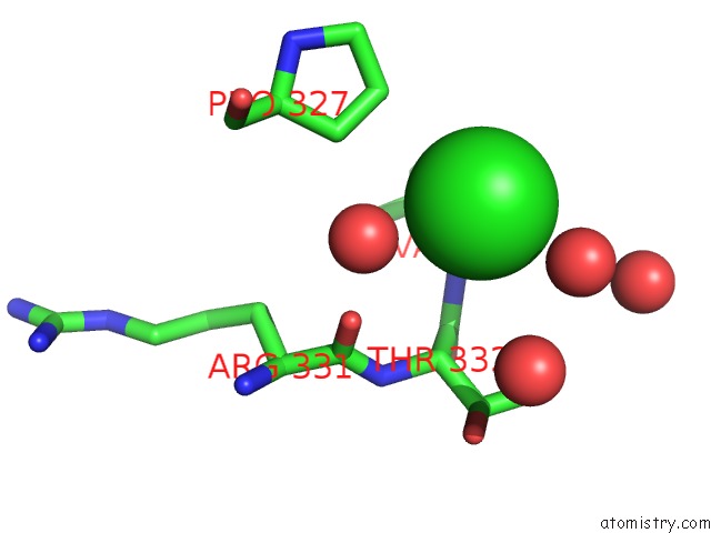
Mono view
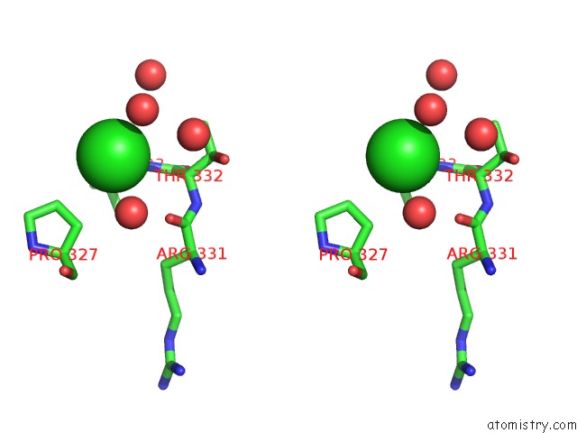
Stereo pair view
Mono view
Stereo pair view
A full contact list of Chlorine with other atoms in the Cl binding
site number 1 of Crystal Structure of Putative Long-Chain-Fatty-Acid Coa Ligase From Rhodopseudomonas Palustris CGA009 within 5.0Å range:
|
Reference:
Y.Patskovsky,
R.Toro,
R.Foti,
M.Dickey,
J.M.Sauder,
S.K.Burley,
S.C.Almo.
Crystal Structure of Putative Long-Chain-Fatty-Acid Coa Synthase From Rhodopseudomonas Palustris CGA009 To Be Published.
Page generated: Fri Jul 11 06:37:24 2025
Last articles
Mg in 5MMJMg in 5MRA
Mg in 5MTV
Mg in 5MS0
Mg in 5MRU
Mg in 5MQJ
Mg in 5MQW
Mg in 5MQT
Mg in 5MQL
Mg in 5MQ1