Chlorine »
PDB 3itd-3jpy »
3iwf »
Chlorine in PDB 3iwf: The Crystal Structure of the N-Terminal Domain of A Rpir Transcriptional Regulator From Staphylococcus Epidermidis to 1.4A
Protein crystallography data
The structure of The Crystal Structure of the N-Terminal Domain of A Rpir Transcriptional Regulator From Staphylococcus Epidermidis to 1.4A, PDB code: 3iwf
was solved by
A.J.Stein,
A.Sather,
M.Borovilos,
M.Bargassa,
A.Joachimiak,
Midwest Centerfor Structural Genomics (Mcsg),
with X-Ray Crystallography technique. A brief refinement statistics is given in the table below:
| Resolution Low / High (Å) | 30.70 / 1.40 |
| Space group | C 2 2 21 |
| Cell size a, b, c (Å), α, β, γ (°) | 48.921, 54.028, 173.044, 90.00, 90.00, 90.00 |
| R / Rfree (%) | 17.7 / 20.5 |
Other elements in 3iwf:
The structure of The Crystal Structure of the N-Terminal Domain of A Rpir Transcriptional Regulator From Staphylococcus Epidermidis to 1.4A also contains other interesting chemical elements:
| Nickel | (Ni) | 2 atoms |
| Sodium | (Na) | 1 atom |
Chlorine Binding Sites:
The binding sites of Chlorine atom in the The Crystal Structure of the N-Terminal Domain of A Rpir Transcriptional Regulator From Staphylococcus Epidermidis to 1.4A
(pdb code 3iwf). This binding sites where shown within
5.0 Angstroms radius around Chlorine atom.
In total 2 binding sites of Chlorine where determined in the The Crystal Structure of the N-Terminal Domain of A Rpir Transcriptional Regulator From Staphylococcus Epidermidis to 1.4A, PDB code: 3iwf:
Jump to Chlorine binding site number: 1; 2;
In total 2 binding sites of Chlorine where determined in the The Crystal Structure of the N-Terminal Domain of A Rpir Transcriptional Regulator From Staphylococcus Epidermidis to 1.4A, PDB code: 3iwf:
Jump to Chlorine binding site number: 1; 2;
Chlorine binding site 1 out of 2 in 3iwf
Go back to
Chlorine binding site 1 out
of 2 in the The Crystal Structure of the N-Terminal Domain of A Rpir Transcriptional Regulator From Staphylococcus Epidermidis to 1.4A
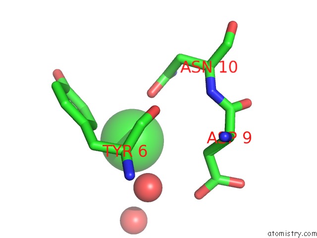
Mono view
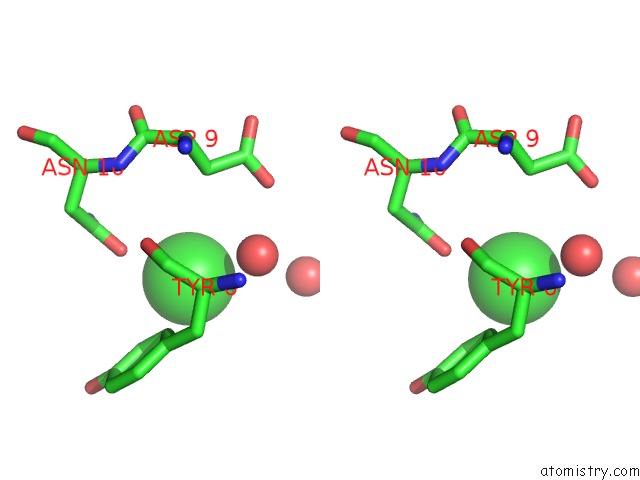
Stereo pair view
Mono view
Stereo pair view
A full contact list of Chlorine with other atoms in the Cl binding
site number 1 of The Crystal Structure of the N-Terminal Domain of A Rpir Transcriptional Regulator From Staphylococcus Epidermidis to 1.4A within 5.0Å range:
|
Chlorine binding site 2 out of 2 in 3iwf
Go back to
Chlorine binding site 2 out
of 2 in the The Crystal Structure of the N-Terminal Domain of A Rpir Transcriptional Regulator From Staphylococcus Epidermidis to 1.4A
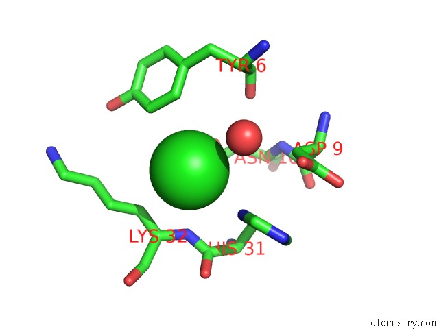
Mono view
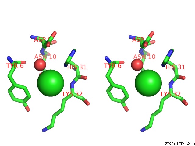
Stereo pair view
Mono view
Stereo pair view
A full contact list of Chlorine with other atoms in the Cl binding
site number 2 of The Crystal Structure of the N-Terminal Domain of A Rpir Transcriptional Regulator From Staphylococcus Epidermidis to 1.4A within 5.0Å range:
|
Reference:
A.J.Stein,
A.Sather,
M.Borovilos,
M.Bargassa,
A.Joachimiak.
The Crystal Structure of the N-Terminal Domain of A Rpir Transcriptional Regulator From Staphylococcus Epidermidis to 1.4A To Be Published.
Page generated: Fri Jul 11 06:37:52 2025
Last articles
K in 5T5MK in 5T30
K in 5T09
K in 5SHY
K in 5SCK
K in 5SCJ
K in 5SCI
K in 5SCH
K in 5SCG
K in 5SCF