Chlorine »
PDB 3jq0-3k1s »
3k1e »
Chlorine in PDB 3k1e: Crystal Structure of Odorant Binding Protein 1 (AAEGOBP1) From Aedes Aegypti
Protein crystallography data
The structure of Crystal Structure of Odorant Binding Protein 1 (AAEGOBP1) From Aedes Aegypti, PDB code: 3k1e
was solved by
N.R.Leite,
R.Krogh,
W.S.Leal,
J.Iulek,
G.Oliva,
with X-Ray Crystallography technique. A brief refinement statistics is given in the table below:
| Resolution Low / High (Å) | 68.68 / 1.85 |
| Space group | P 1 21 1 |
| Cell size a, b, c (Å), α, β, γ (°) | 34.248, 47.870, 69.082, 90.00, 96.61, 90.00 |
| R / Rfree (%) | 15.1 / 21.2 |
Other elements in 3k1e:
The structure of Crystal Structure of Odorant Binding Protein 1 (AAEGOBP1) From Aedes Aegypti also contains other interesting chemical elements:
| Magnesium | (Mg) | 3 atoms |
Chlorine Binding Sites:
The binding sites of Chlorine atom in the Crystal Structure of Odorant Binding Protein 1 (AAEGOBP1) From Aedes Aegypti
(pdb code 3k1e). This binding sites where shown within
5.0 Angstroms radius around Chlorine atom.
In total only one binding site of Chlorine was determined in the Crystal Structure of Odorant Binding Protein 1 (AAEGOBP1) From Aedes Aegypti, PDB code: 3k1e:
In total only one binding site of Chlorine was determined in the Crystal Structure of Odorant Binding Protein 1 (AAEGOBP1) From Aedes Aegypti, PDB code: 3k1e:
Chlorine binding site 1 out of 1 in 3k1e
Go back to
Chlorine binding site 1 out
of 1 in the Crystal Structure of Odorant Binding Protein 1 (AAEGOBP1) From Aedes Aegypti
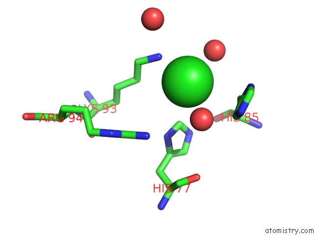
Mono view
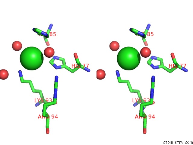
Stereo pair view
Mono view
Stereo pair view
A full contact list of Chlorine with other atoms in the Cl binding
site number 1 of Crystal Structure of Odorant Binding Protein 1 (AAEGOBP1) From Aedes Aegypti within 5.0Å range:
|
Reference:
N.R.Leite,
R.Krogh,
W.Xu,
Y.Ishida,
J.Iulek,
W.S.Leal,
G.Oliva.
Structure of An Odorant-Binding Protein From the Mosquito Aedes Aegypti Suggests A Binding Pocket Covered By A pH-Sensitive "Lid". Plos One V. 4 E8006 2009.
ISSN: ESSN 1932-6203
PubMed: 19956631
DOI: 10.1371/JOURNAL.PONE.0008006
Page generated: Fri Jul 11 06:48:33 2025
ISSN: ESSN 1932-6203
PubMed: 19956631
DOI: 10.1371/JOURNAL.PONE.0008006
Last articles
Mg in 3CR1Mg in 3CME
Mg in 3CMA
Mg in 3CP6
Mg in 3CPJ
Mg in 3CPH
Mg in 3CON
Mg in 3CMV
Mg in 3COB
Mg in 3CNZ