Chlorine »
PDB 3k1t-3k9t »
3k2o »
Chlorine in PDB 3k2o: Structure of An Oxygenase
Protein crystallography data
The structure of Structure of An Oxygenase, PDB code: 3k2o
was solved by
T.Krojer,
M.A.Mcdonough,
I.J.Clifton,
M.Mantri,
S.S.Ng,
A.C.W.Pike,
D.S.Butler,
C.J.Webby,
G.Kochan,
C.Bhatia,
J.E.Bray,
A.Chaikuad,
O.Gileadi,
F.Von Delft,
J.Weigelt,
C.H.Arrowsmith,
C.Bountra,
A.M.Edwards,
C.J.Schofield,
K.L.Kavanagh,
U.Oppermann,
with X-Ray Crystallography technique. A brief refinement statistics is given in the table below:
| Resolution Low / High (Å) | 19.85 / 1.75 |
| Space group | P 1 21 1 |
| Cell size a, b, c (Å), α, β, γ (°) | 49.440, 102.030, 98.218, 90.00, 95.89, 90.00 |
| R / Rfree (%) | 19.2 / 22.5 |
Other elements in 3k2o:
The structure of Structure of An Oxygenase also contains other interesting chemical elements:
| Nickel | (Ni) | 2 atoms |
| Sodium | (Na) | 2 atoms |
Chlorine Binding Sites:
The binding sites of Chlorine atom in the Structure of An Oxygenase
(pdb code 3k2o). This binding sites where shown within
5.0 Angstroms radius around Chlorine atom.
In total 6 binding sites of Chlorine where determined in the Structure of An Oxygenase, PDB code: 3k2o:
Jump to Chlorine binding site number: 1; 2; 3; 4; 5; 6;
In total 6 binding sites of Chlorine where determined in the Structure of An Oxygenase, PDB code: 3k2o:
Jump to Chlorine binding site number: 1; 2; 3; 4; 5; 6;
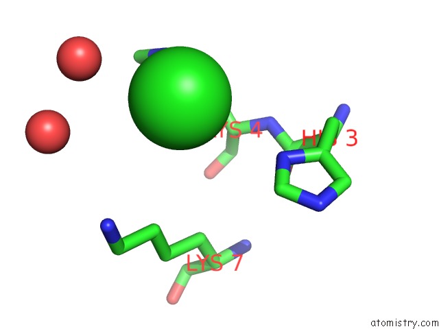
Chlorine binding site 1 out of 6 in 3k2o
Go back to
Chlorine binding site 1 out
of 6 in the Structure of An Oxygenase

Mono view

Stereo pair view
Mono view
Stereo pair view
A full contact list of Chlorine with other atoms in the Cl binding
site number 1 of Structure of An Oxygenase within 5.0Å range:
|
Chlorine binding site 2 out of 6 in 3k2o
Go back to
Chlorine binding site 2 out
of 6 in the Structure of An Oxygenase
Mono view

Stereo pair view
Mono view
Stereo pair view
A full contact list of Chlorine with other atoms in the Cl binding
site number 2 of Structure of An Oxygenase within 5.0Å range:
|
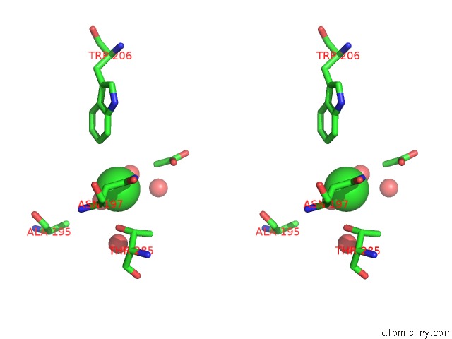
Chlorine binding site 3 out of 6 in 3k2o
Go back to
Chlorine binding site 3 out
of 6 in the Structure of An Oxygenase

Mono view
Stereo pair view
Mono view
Stereo pair view
A full contact list of Chlorine with other atoms in the Cl binding
site number 3 of Structure of An Oxygenase within 5.0Å range:
|
Chlorine binding site 4 out of 6 in 3k2o
Go back to
Chlorine binding site 4 out
of 6 in the Structure of An Oxygenase

Mono view

Stereo pair view
Mono view
Stereo pair view
A full contact list of Chlorine with other atoms in the Cl binding
site number 4 of Structure of An Oxygenase within 5.0Å range:
|
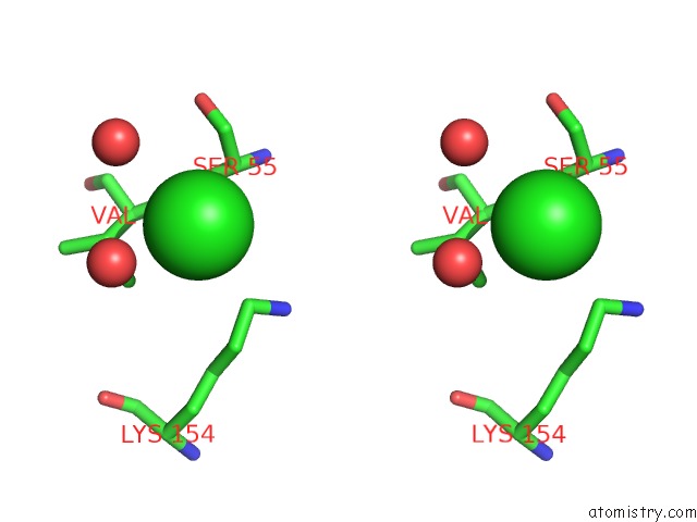
Chlorine binding site 5 out of 6 in 3k2o
Go back to
Chlorine binding site 5 out
of 6 in the Structure of An Oxygenase

Mono view
Stereo pair view
Mono view
Stereo pair view
A full contact list of Chlorine with other atoms in the Cl binding
site number 5 of Structure of An Oxygenase within 5.0Å range:
|
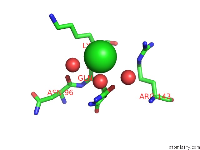
Chlorine binding site 6 out of 6 in 3k2o
Go back to
Chlorine binding site 6 out
of 6 in the Structure of An Oxygenase
Mono view

Stereo pair view
Mono view
Stereo pair view
A full contact list of Chlorine with other atoms in the Cl binding
site number 6 of Structure of An Oxygenase within 5.0Å range:
|
Reference:
M.Mantri,
T.Krojer,
E.A.Bagg,
C.A.Webby,
D.S.Butler,
G.Kochan,
K.L.Kavanagh,
U.Oppermann,
M.A.Mcdonough,
C.J.Schofield.
Crystal Structure of the 2-Oxoglutarate- and Fe(II)-Dependent Lysyl Hydroxylase JMJD6. J.Mol.Biol. V. 401 211 2010.
ISSN: ISSN 0022-2836
PubMed: 20685276
DOI: 10.1016/J.JMB.2010.05.054
Page generated: Fri Jul 11 06:49:59 2025
ISSN: ISSN 0022-2836
PubMed: 20685276
DOI: 10.1016/J.JMB.2010.05.054
Last articles
K in 1D7UK in 1D7S
K in 1D7R
K in 1C38
K in 1CPG
K in 1CPE
K in 1C30
K in 1C7J
K in 1C35
K in 1BXR