Chlorine »
PDB 3k9u-3khi »
3kh2 »
Chlorine in PDB 3kh2: Crystal Structure of the P1 Bacteriophage Doc Toxin (F68S) in Complex with the Phd Antitoxin (L17M/V39A). Northeast Structural Genomics Targets ER385-ER386
Protein crystallography data
The structure of Crystal Structure of the P1 Bacteriophage Doc Toxin (F68S) in Complex with the Phd Antitoxin (L17M/V39A). Northeast Structural Genomics Targets ER385-ER386, PDB code: 3kh2
was solved by
M.A.Arbing,
A.P.Kuzin,
M.Su,
M.Abashidze,
G.Verdon,
M.Liu,
R.Xiao,
T.Acton,
M.Inouye,
G.T.Montelione,
N.A.Woychik,
J.F.Hunt,
Northeast Structuralgenomics Consortium (Nesg),
with X-Ray Crystallography technique. A brief refinement statistics is given in the table below:
| Resolution Low / High (Å) | 30.00 / 2.71 |
| Space group | P 21 21 21 |
| Cell size a, b, c (Å), α, β, γ (°) | 95.899, 111.277, 118.747, 90.00, 90.00, 90.00 |
| R / Rfree (%) | 21.7 / 26.7 |
Chlorine Binding Sites:
The binding sites of Chlorine atom in the Crystal Structure of the P1 Bacteriophage Doc Toxin (F68S) in Complex with the Phd Antitoxin (L17M/V39A). Northeast Structural Genomics Targets ER385-ER386
(pdb code 3kh2). This binding sites where shown within
5.0 Angstroms radius around Chlorine atom.
In total only one binding site of Chlorine was determined in the Crystal Structure of the P1 Bacteriophage Doc Toxin (F68S) in Complex with the Phd Antitoxin (L17M/V39A). Northeast Structural Genomics Targets ER385-ER386, PDB code: 3kh2:
In total only one binding site of Chlorine was determined in the Crystal Structure of the P1 Bacteriophage Doc Toxin (F68S) in Complex with the Phd Antitoxin (L17M/V39A). Northeast Structural Genomics Targets ER385-ER386, PDB code: 3kh2:
Chlorine binding site 1 out of 1 in 3kh2
Go back to
Chlorine binding site 1 out
of 1 in the Crystal Structure of the P1 Bacteriophage Doc Toxin (F68S) in Complex with the Phd Antitoxin (L17M/V39A). Northeast Structural Genomics Targets ER385-ER386
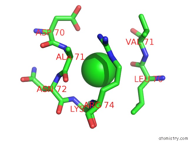
Mono view
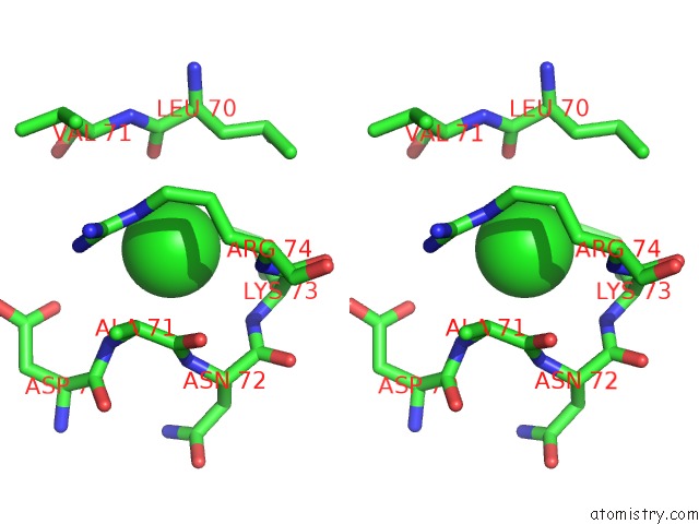
Stereo pair view
Mono view
Stereo pair view
A full contact list of Chlorine with other atoms in the Cl binding
site number 1 of Crystal Structure of the P1 Bacteriophage Doc Toxin (F68S) in Complex with the Phd Antitoxin (L17M/V39A). Northeast Structural Genomics Targets ER385-ER386 within 5.0Å range:
|
Reference:
M.A.Arbing,
S.K.Handelman,
A.P.Kuzin,
G.Verdon,
C.Wang,
M.Su,
F.P.Rothenbacher,
M.Abashidze,
M.Liu,
J.M.Hurley,
R.Xiao,
T.Acton,
M.Inouye,
G.T.Montelione,
N.A.Woychik,
J.F.Hunt.
Crystal Structures of Phd-Doc, Higa, and Yeeu Establish Multiple Evolutionary Links Between Microbial Growth-Regulating Toxin-Antitoxin Systems. Structure V. 18 996 2010.
ISSN: ISSN 0969-2126
PubMed: 20696400
DOI: 10.1016/J.STR.2010.04.018
Page generated: Fri Jul 11 07:00:09 2025
ISSN: ISSN 0969-2126
PubMed: 20696400
DOI: 10.1016/J.STR.2010.04.018
Last articles
Zn in 9QM9Zn in 9S44
Zn in 9OFE
Zn in 9OFC
Zn in 9OFD
Zn in 9OF1
Zn in 9OFB
Zn in 9N0J
Zn in 9M5X
Zn in 9LGI