Chlorine »
PDB 3khp-3ktf »
3kia »
Chlorine in PDB 3kia: Crystal Structure of Mannosyl-3-Phosphoglycerate Synthase From Rubrobacter Xylanophilus
Enzymatic activity of Crystal Structure of Mannosyl-3-Phosphoglycerate Synthase From Rubrobacter Xylanophilus
All present enzymatic activity of Crystal Structure of Mannosyl-3-Phosphoglycerate Synthase From Rubrobacter Xylanophilus:
2.4.1.217;
2.4.1.217;
Protein crystallography data
The structure of Crystal Structure of Mannosyl-3-Phosphoglycerate Synthase From Rubrobacter Xylanophilus, PDB code: 3kia
was solved by
S.Macedo-Ribeiro,
P.J.B.Pereira,
N.Empadinhas,
M.S.Da Costa,
with X-Ray Crystallography technique. A brief refinement statistics is given in the table below:
| Resolution Low / High (Å) | 53.70 / 2.80 |
| Space group | P 65 2 2 |
| Cell size a, b, c (Å), α, β, γ (°) | 109.010, 109.010, 313.418, 90.00, 90.00, 120.00 |
| R / Rfree (%) | 19 / 23.9 |
Other elements in 3kia:
The structure of Crystal Structure of Mannosyl-3-Phosphoglycerate Synthase From Rubrobacter Xylanophilus also contains other interesting chemical elements:
| Magnesium | (Mg) | 3 atoms |
Chlorine Binding Sites:
The binding sites of Chlorine atom in the Crystal Structure of Mannosyl-3-Phosphoglycerate Synthase From Rubrobacter Xylanophilus
(pdb code 3kia). This binding sites where shown within
5.0 Angstroms radius around Chlorine atom.
In total 3 binding sites of Chlorine where determined in the Crystal Structure of Mannosyl-3-Phosphoglycerate Synthase From Rubrobacter Xylanophilus, PDB code: 3kia:
Jump to Chlorine binding site number: 1; 2; 3;
In total 3 binding sites of Chlorine where determined in the Crystal Structure of Mannosyl-3-Phosphoglycerate Synthase From Rubrobacter Xylanophilus, PDB code: 3kia:
Jump to Chlorine binding site number: 1; 2; 3;
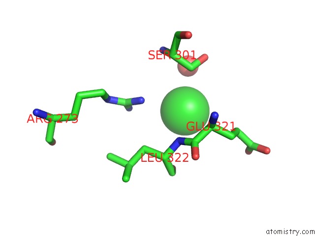
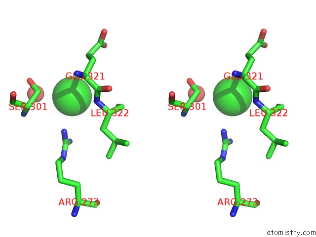
Chlorine binding site 1 out of 3 in 3kia
Go back to
Chlorine binding site 1 out
of 3 in the Crystal Structure of Mannosyl-3-Phosphoglycerate Synthase From Rubrobacter Xylanophilus
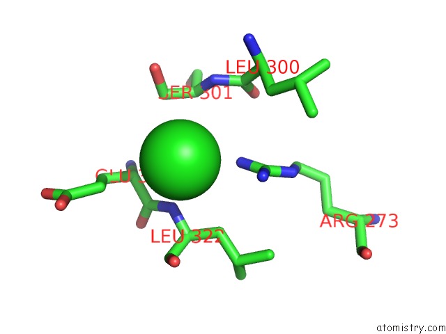
Mono view
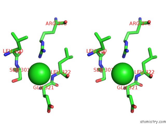
Stereo pair view
Mono view
Stereo pair view
A full contact list of Chlorine with other atoms in the Cl binding
site number 1 of Crystal Structure of Mannosyl-3-Phosphoglycerate Synthase From Rubrobacter Xylanophilus within 5.0Å range:
|
Chlorine binding site 2 out of 3 in 3kia
Go back to
Chlorine binding site 2 out
of 3 in the Crystal Structure of Mannosyl-3-Phosphoglycerate Synthase From Rubrobacter Xylanophilus
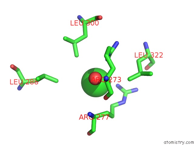
Mono view
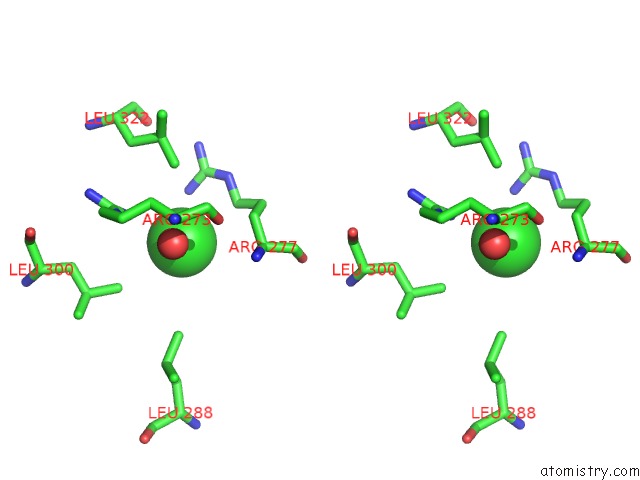
Stereo pair view
Mono view
Stereo pair view
A full contact list of Chlorine with other atoms in the Cl binding
site number 2 of Crystal Structure of Mannosyl-3-Phosphoglycerate Synthase From Rubrobacter Xylanophilus within 5.0Å range:
|
Chlorine binding site 3 out of 3 in 3kia
Go back to
Chlorine binding site 3 out
of 3 in the Crystal Structure of Mannosyl-3-Phosphoglycerate Synthase From Rubrobacter Xylanophilus
Mono view
Stereo pair view
Mono view
Stereo pair view
A full contact list of Chlorine with other atoms in the Cl binding
site number 3 of Crystal Structure of Mannosyl-3-Phosphoglycerate Synthase From Rubrobacter Xylanophilus within 5.0Å range:
|
Reference:
N.Empadinhas,
P.J.B.Pereira,
L.Albuquerque,
J.Costa,
B.Sa-Moura,
A.T.Marques,
S.Macedo-Ribeiro,
M.S.Da Costa.
Functional and Structural Characterization of A Novel Mannosyl-3-Phosphoglycerate Synthase From Rubrobacter Xylanophilus Reveals Its Dual Substrate Specificity Mol.Microbiol. V. 79 76 2011.
ISSN: ISSN 0950-382X
PubMed: 21166895
DOI: 10.1111/J.1365-2958.2010.07432.X
Page generated: Fri Jul 11 07:00:35 2025
ISSN: ISSN 0950-382X
PubMed: 21166895
DOI: 10.1111/J.1365-2958.2010.07432.X
Last articles
K in 8X9OK in 8XAK
K in 8X9M
K in 8X70
K in 8WUW
K in 8WUX
K in 8X0S
K in 8X4R
K in 8WGR
K in 8W9V