Chlorine »
PDB 3khp-3ktf »
3kka »
Chlorine in PDB 3kka: Co-Crystal Structure of the Sam Domains of EPHA1 and EPHA2
Enzymatic activity of Co-Crystal Structure of the Sam Domains of EPHA1 and EPHA2
All present enzymatic activity of Co-Crystal Structure of the Sam Domains of EPHA1 and EPHA2:
2.7.10.1;
2.7.10.1;
Protein crystallography data
The structure of Co-Crystal Structure of the Sam Domains of EPHA1 and EPHA2, PDB code: 3kka
was solved by
J.R.Walker,
L.Yermekbayeva,
C.Butler-Cole,
J.Weigelt,
C.Bountra,
C.H.Arrowsmith,
A.M.Edwards,
A.Bochkarev,
S.Dhe-Paganon,
Structural Genomics Consortium (Sgc),
with X-Ray Crystallography technique. A brief refinement statistics is given in the table below:
| Resolution Low / High (Å) | 30.44 / 2.40 |
| Space group | P 21 21 21 |
| Cell size a, b, c (Å), α, β, γ (°) | 57.554, 56.071, 107.617, 90.00, 90.00, 90.00 |
| R / Rfree (%) | 20 / 23.5 |
Chlorine Binding Sites:
The binding sites of Chlorine atom in the Co-Crystal Structure of the Sam Domains of EPHA1 and EPHA2
(pdb code 3kka). This binding sites where shown within
5.0 Angstroms radius around Chlorine atom.
In total only one binding site of Chlorine was determined in the Co-Crystal Structure of the Sam Domains of EPHA1 and EPHA2, PDB code: 3kka:
In total only one binding site of Chlorine was determined in the Co-Crystal Structure of the Sam Domains of EPHA1 and EPHA2, PDB code: 3kka:
Chlorine binding site 1 out of 1 in 3kka
Go back to
Chlorine binding site 1 out
of 1 in the Co-Crystal Structure of the Sam Domains of EPHA1 and EPHA2
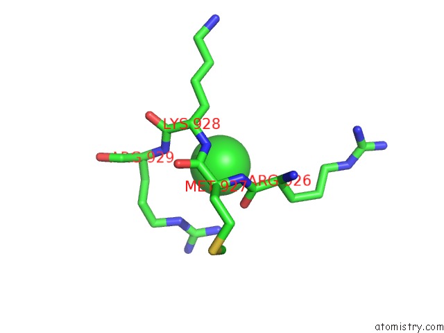
Mono view
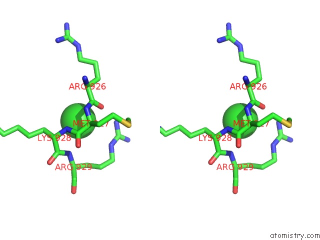
Stereo pair view
Mono view
Stereo pair view
A full contact list of Chlorine with other atoms in the Cl binding
site number 1 of Co-Crystal Structure of the Sam Domains of EPHA1 and EPHA2 within 5.0Å range:
|
Reference:
J.R.Walker,
L.Yermekbayeva,
C.Butler-Cole,
J.Weigelt,
C.Bountra,
C.H.Arrowsmith,
A.M.Edwards,
A.Bochkarev,
S.Dhe-Paganon.
Co-Crystal Structure of the Sam Domains of Human Ephrin Type-A Receptor 1 and Human Ephrin Type-A Receptor 2 To Be Published.
Page generated: Fri Jul 11 07:01:10 2025
Last articles
Zn in 3N6FZn in 3N6E
Zn in 3N6D
Zn in 3N6C
Zn in 3N6B
Zn in 3N66
Zn in 3N6A
Zn in 3N69
Zn in 3N68
Zn in 3N65