Atomistry » Chlorine » PDB 3ku9-3l29 » 3kxp
Atomistry »
  Chlorine »
    PDB 3ku9-3l29 »
      3kxp »

Chlorine in PDB 3kxp: Crystal Structure of E-2-(Acetamidomethylene)Succinate Hydrolase

Protein crystallography data

The structure of Crystal Structure of E-2-(Acetamidomethylene)Succinate Hydrolase, PDB code: 3kxp was solved by K.M.Mcculloch, T.Mukherjee, T.P.Begley, S.E.Ealick, with X-Ray Crystallography technique. A brief refinement statistics is given in the table below:

Resolution Low / High (Å) 50.00 / 2.26
Space group P 21 21 21
Cell size a, b, c (Å), α, β, γ (°) 115.200, 178.530, 189.250, 90.00, 90.00, 90.00
R / Rfree (%) 20.4 / 24.5

Chlorine Binding Sites:

Pages:

>>> Page 1 <<< Page 2, Binding sites: 11 - 12;

Binding sites:

The binding sites of Chlorine atom in the Crystal Structure of E-2-(Acetamidomethylene)Succinate Hydrolase (pdb code 3kxp). This binding sites where shown within 5.0 Angstroms radius around Chlorine atom.
In total 12 binding sites of Chlorine where determined in the Crystal Structure of E-2-(Acetamidomethylene)Succinate Hydrolase, PDB code: 3kxp:
Jump to Chlorine binding site number: 1; 2; 3; 4; 5; 6; 7; 8; 9; 10;

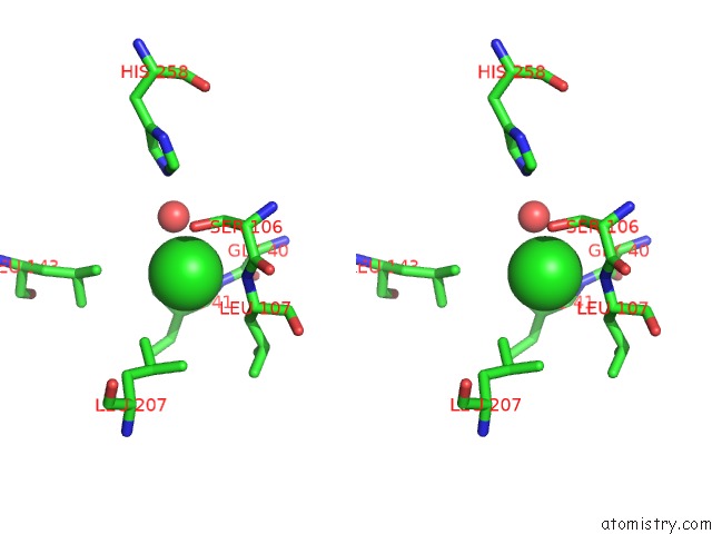
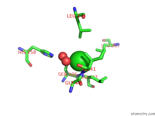
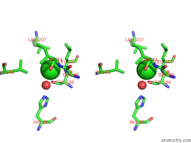
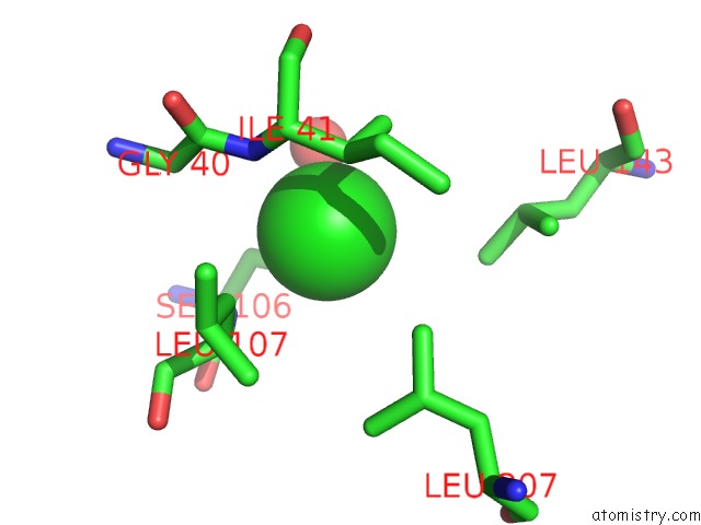
Chlorine binding site 1 out of 12 in 3kxp

Go back to Chlorine Binding Sites List in 3kxp
Chlorine binding site 1 out of 12 in the Crystal Structure of E-2-(Acetamidomethylene)Succinate Hydrolase


Mono view


Stereo pair view

A full contact list of Chlorine with other atoms in the Cl binding site number 1 of Crystal Structure of E-2-(Acetamidomethylene)Succinate Hydrolase within 5.0Å range:
probe atom residue distance (Å) B Occ
A:Cl279

b:25.8
occ:1.00
OG A:SER106 2.9 19.1 1.0
O A:HOH630 3.0 30.2 1.0
N A:ILE41 3.2 16.4 1.0
N A:LEU107 3.3 23.6 1.0
CB A:SER106 3.4 19.0 1.0
CG2 A:ILE41 3.5 19.4 1.0
CB A:ILE41 3.5 17.6 1.0
CB A:LEU107 3.6 22.0 1.0
CA A:LEU107 3.9 21.9 1.0
CD1 A:LEU207 3.9 23.5 1.0
CA A:ILE41 4.0 17.8 1.0
C A:GLY40 4.2 18.1 1.0
CA A:GLY40 4.2 16.3 1.0
CD1 A:LEU143 4.2 32.8 1.0
C A:SER106 4.2 22.8 1.0
CA A:SER106 4.4 21.4 1.0
NE2 A:HIS258 4.9 23.5 1.0
CG1 A:ILE41 4.9 19.1 1.0

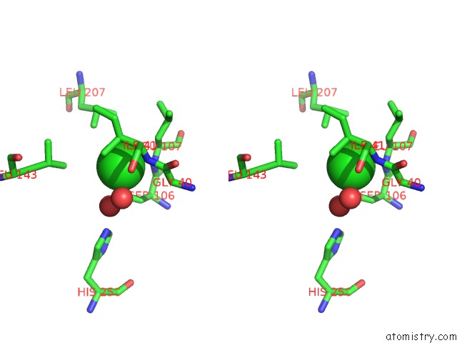
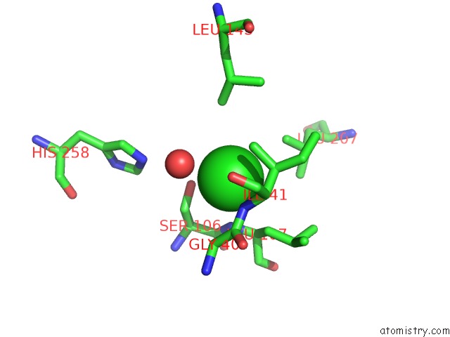
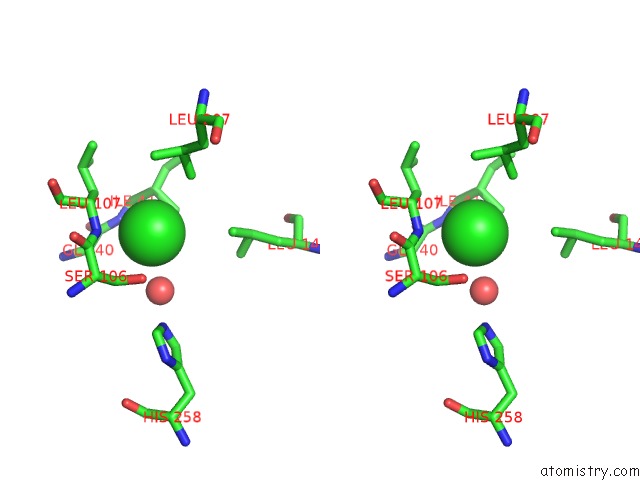
Chlorine binding site 2 out of 12 in 3kxp

Go back to Chlorine Binding Sites List in 3kxp
Chlorine binding site 2 out of 12 in the Crystal Structure of E-2-(Acetamidomethylene)Succinate Hydrolase


Mono view


Stereo pair view

A full contact list of Chlorine with other atoms in the Cl binding site number 2 of Crystal Structure of E-2-(Acetamidomethylene)Succinate Hydrolase within 5.0Å range:
probe atom residue distance (Å) B Occ
B:Cl279

b:33.6
occ:1.00
O B:HOH455 2.8 29.2 1.0
OG B:SER106 3.1 28.2 1.0
N B:LEU107 3.2 24.7 1.0
N B:ILE41 3.3 28.0 1.0
CB B:SER106 3.4 26.2 1.0
CG2 B:ILE41 3.4 25.7 1.0
CB B:LEU107 3.5 24.3 1.0
CB B:ILE41 3.6 26.9 1.0
CA B:LEU107 3.8 25.3 1.0
CD1 B:LEU207 3.9 34.6 1.0
CA B:ILE41 4.1 25.7 1.0
CA B:GLY40 4.2 27.6 1.0
C B:SER106 4.2 25.9 1.0
CD1 B:LEU143 4.2 42.5 1.0
C B:GLY40 4.2 29.3 1.0
CA B:SER106 4.4 25.9 1.0
CD2 B:LEU207 4.8 36.4 1.0
CG B:LEU107 4.8 23.7 1.0
CG1 B:ILE41 5.0 26.9 1.0
CG B:LEU207 5.0 36.2 1.0

Chlorine binding site 3 out of 12 in 3kxp

Go back to Chlorine Binding Sites List in 3kxp
Chlorine binding site 3 out of 12 in the Crystal Structure of E-2-(Acetamidomethylene)Succinate Hydrolase


Mono view


Stereo pair view

A full contact list of Chlorine with other atoms in the Cl binding site number 3 of Crystal Structure of E-2-(Acetamidomethylene)Succinate Hydrolase within 5.0Å range:
probe atom residue distance (Å) B Occ
C:Cl279

b:26.8
occ:1.00
OG C:SER106 2.9 24.6 1.0
O C:HOH737 3.1 26.5 1.0
N C:ILE41 3.1 22.2 1.0
N C:LEU107 3.2 19.3 1.0
CG2 C:ILE41 3.3 21.0 1.0
CB C:SER106 3.4 22.9 1.0
CB C:ILE41 3.5 20.1 1.0
CB C:LEU107 3.5 20.2 1.0
CA C:LEU107 3.8 21.9 1.0
CD1 C:LEU207 3.8 23.5 1.0
CA C:ILE41 3.9 22.0 1.0
O C:HOH728 3.9 49.9 1.0
C C:GLY40 4.1 25.1 1.0
CA C:GLY40 4.1 25.8 1.0
C C:SER106 4.1 20.0 1.0
CD1 C:LEU143 4.2 38.2 1.0
CA C:SER106 4.4 21.2 1.0
CG C:LEU107 4.9 22.6 1.0
CG1 C:ILE41 4.9 20.4 1.0
NE2 C:HIS258 4.9 25.5 1.0

Chlorine binding site 4 out of 12 in 3kxp

Go back to Chlorine Binding Sites List in 3kxp
Chlorine binding site 4 out of 12 in the Crystal Structure of E-2-(Acetamidomethylene)Succinate Hydrolase


Mono view


Stereo pair view

A full contact list of Chlorine with other atoms in the Cl binding site number 4 of Crystal Structure of E-2-(Acetamidomethylene)Succinate Hydrolase within 5.0Å range:
probe atom residue distance (Å) B Occ
D:Cl279

b:35.8
occ:1.00
OG D:SER106 2.8 32.4 1.0
O D:HOH339 3.1 33.2 1.0
N D:LEU107 3.2 22.9 1.0
N D:ILE41 3.2 25.2 1.0
CG2 D:ILE41 3.3 24.7 1.0
CB D:SER106 3.3 28.5 1.0
CB D:ILE41 3.5 23.6 1.0
CB D:LEU107 3.6 25.2 1.0
CA D:LEU107 3.8 25.2 1.0
CA D:ILE41 4.0 25.1 1.0
CD1 D:LEU207 4.1 29.7 1.0
C D:SER106 4.1 25.0 1.0
O D:HOH1084 4.1 71.4 1.0
C D:GLY40 4.2 25.6 1.0
CA D:GLY40 4.2 26.0 1.0
CD1 D:LEU143 4.2 32.9 1.0
CA D:SER106 4.4 27.1 1.0
CD2 D:LEU207 4.7 28.5 1.0
NE2 D:HIS258 4.8 23.7 1.0
CG1 D:ILE41 4.9 25.7 1.0
CG D:LEU107 5.0 25.4 1.0

Chlorine binding site 5 out of 12 in 3kxp

Go back to Chlorine Binding Sites List in 3kxp
Chlorine binding site 5 out of 12 in the Crystal Structure of E-2-(Acetamidomethylene)Succinate Hydrolase


Mono view


Stereo pair view

A full contact list of Chlorine with other atoms in the Cl binding site number 5 of Crystal Structure of E-2-(Acetamidomethylene)Succinate Hydrolase within 5.0Å range:
probe atom residue distance (Å) B Occ
E:Cl279

b:33.4
occ:1.00
OG E:SER106 2.9 31.7 1.0
O E:HOH794 3.0 34.9 1.0
N E:ILE41 3.1 29.3 1.0
N E:LEU107 3.2 27.4 1.0
CG2 E:ILE41 3.4 28.7 1.0
CB E:SER106 3.4 26.1 1.0
CB E:LEU107 3.4 29.0 1.0
CB E:ILE41 3.5 27.3 1.0
CA E:LEU107 3.8 28.2 1.0
CA E:ILE41 3.9 29.7 1.0
C E:GLY40 4.0 29.4 1.0
CA E:GLY40 4.0 29.6 1.0
CD1 E:LEU207 4.0 37.1 1.0
C E:SER106 4.2 27.1 1.0
CD1 E:LEU143 4.2 44.9 1.0
CA E:SER106 4.4 27.7 1.0
CG E:LEU107 4.8 27.8 1.0
CD2 E:LEU207 4.9 35.9 1.0
NE2 E:HIS258 4.9 36.7 1.0
CG1 E:ILE41 5.0 30.0 1.0

Chlorine binding site 6 out of 12 in 3kxp

Go back to Chlorine Binding Sites List in 3kxp
Chlorine binding site 6 out of 12 in the Crystal Structure of E-2-(Acetamidomethylene)Succinate Hydrolase


Mono view


Stereo pair view

A full contact list of Chlorine with other atoms in the Cl binding site number 6 of Crystal Structure of E-2-(Acetamidomethylene)Succinate Hydrolase within 5.0Å range:
probe atom residue distance (Å) B Occ
F:Cl279

b:32.8
occ:1.00
OG F:SER106 3.0 29.1 1.0
O F:HOH432 3.1 38.6 1.0
CG2 F:ILE41 3.2 24.2 1.0
N F:ILE41 3.2 23.8 1.0
N F:LEU107 3.2 31.4 1.0
CB F:ILE41 3.4 24.5 1.0
CB F:LEU107 3.5 31.7 1.0
CB F:SER106 3.5 29.1 1.0
CD1 F:LEU207 3.7 37.7 1.0
CA F:LEU107 3.8 32.1 1.0
CA F:ILE41 3.9 24.3 1.0
CD1 F:LEU143 4.1 36.6 1.0
C F:GLY40 4.2 25.2 1.0
CA F:GLY40 4.2 26.3 1.0
C F:SER106 4.2 30.9 1.0
CA F:SER106 4.5 30.7 1.0
CG F:LEU107 4.8 31.5 1.0
CG1 F:ILE41 4.8 23.2 1.0

Chlorine binding site 7 out of 12 in 3kxp

Go back to Chlorine Binding Sites List in 3kxp
Chlorine binding site 7 out of 12 in the Crystal Structure of E-2-(Acetamidomethylene)Succinate Hydrolase


Mono view


Stereo pair view

A full contact list of Chlorine with other atoms in the Cl binding site number 7 of Crystal Structure of E-2-(Acetamidomethylene)Succinate Hydrolase within 5.0Å range:
probe atom residue distance (Å) B Occ
G:Cl279

b:31.2
occ:1.00
OG G:SER106 2.7 35.7 1.0
O G:HOH375 3.0 30.1 1.0
N G:ILE41 3.2 22.9 1.0
N G:LEU107 3.3 24.8 1.0
CG2 G:ILE41 3.4 19.1 1.0
CB G:SER106 3.4 28.4 1.0
CB G:ILE41 3.4 19.6 1.0
CB G:LEU107 3.6 22.6 1.0
CA G:LEU107 3.9 24.1 1.0
CD1 G:LEU207 3.9 29.3 1.0
CA G:ILE41 3.9 19.8 1.0
C G:GLY40 4.1 24.2 1.0
CA G:GLY40 4.2 24.2 1.0
CD1 G:LEU143 4.3 37.5 1.0
C G:SER106 4.3 28.3 1.0
CA G:SER106 4.5 28.7 1.0
CG1 G:ILE41 4.8 21.3 1.0
CG G:LEU107 4.9 20.7 1.0

Chlorine binding site 8 out of 12 in 3kxp

Go back to Chlorine Binding Sites List in 3kxp
Chlorine binding site 8 out of 12 in the Crystal Structure of E-2-(Acetamidomethylene)Succinate Hydrolase


Mono view


Stereo pair view

A full contact list of Chlorine with other atoms in the Cl binding site number 8 of Crystal Structure of E-2-(Acetamidomethylene)Succinate Hydrolase within 5.0Å range:
probe atom residue distance (Å) B Occ
H:Cl279

b:29.9
occ:1.00
OG H:SER106 2.9 27.0 1.0
O H:HOH293 3.1 23.4 1.0
CG2 H:ILE41 3.2 18.2 1.0
N H:ILE41 3.2 21.7 1.0
N H:LEU107 3.3 24.6 1.0
CB H:SER106 3.5 26.3 1.0
CB H:ILE41 3.5 18.9 1.0
CB H:LEU107 3.6 21.1 1.0
CA H:LEU107 3.8 24.1 1.0
CD1 H:LEU207 3.8 25.9 1.0
CA H:ILE41 4.0 20.6 1.0
CA H:GLY40 4.1 25.2 1.0
C H:GLY40 4.1 25.3 1.0
C H:SER106 4.2 25.3 1.0
CD2 H:LEU143 4.3 26.1 1.0
CA H:SER106 4.5 25.6 1.0
CD2 H:LEU207 4.8 22.1 1.0
CG H:LEU107 4.9 18.9 1.0
CG1 H:ILE41 4.9 17.7 1.0
CG H:LEU207 5.0 25.6 1.0
CD1 H:LEU143 5.0 28.6 1.0

Chlorine binding site 9 out of 12 in 3kxp

Go back to Chlorine Binding Sites List in 3kxp
Chlorine binding site 9 out of 12 in the Crystal Structure of E-2-(Acetamidomethylene)Succinate Hydrolase


Mono view


Stereo pair view

A full contact list of Chlorine with other atoms in the Cl binding site number 9 of Crystal Structure of E-2-(Acetamidomethylene)Succinate Hydrolase within 5.0Å range:
probe atom residue distance (Å) B Occ
I:Cl279

b:23.0
occ:1.00
CG2 I:ILE41 3.1 16.4 1.0
OG I:SER106 3.1 21.8 1.0
N I:ILE41 3.3 21.1 1.0
N I:LEU107 3.3 19.0 1.0
O I:HOH321 3.3 34.0 1.0
CB I:SER106 3.5 21.5 1.0
CB I:ILE41 3.6 19.1 1.0
CB I:LEU107 3.6 18.9 1.0
CA I:LEU107 3.9 19.2 1.0
CD1 I:LEU207 3.9 29.1 1.0
CA I:ILE41 4.1 20.2 1.0
CA I:GLY40 4.1 20.0 1.0
C I:GLY40 4.2 21.6 1.0
CD2 I:LEU143 4.3 28.9 1.0
C I:SER106 4.3 19.8 1.0
CA I:SER106 4.5 21.4 1.0
CD2 I:LEU207 4.9 26.0 1.0
NE2 I:HIS258 4.9 29.3 1.0
CG I:LEU107 5.0 17.2 1.0
CG1 I:ILE41 5.0 19.5 1.0

Chlorine binding site 10 out of 12 in 3kxp

Go back to Chlorine Binding Sites List in 3kxp
Chlorine binding site 10 out of 12 in the Crystal Structure of E-2-(Acetamidomethylene)Succinate Hydrolase


Mono view


Stereo pair view

A full contact list of Chlorine with other atoms in the Cl binding site number 10 of Crystal Structure of E-2-(Acetamidomethylene)Succinate Hydrolase within 5.0Å range:
probe atom residue distance (Å) B Occ
J:Cl279

b:25.2
occ:1.00
OG J:SER106 2.9 19.9 1.0
N J:ILE41 3.2 22.0 1.0
N J:LEU107 3.3 25.1 1.0
CB J:SER106 3.4 21.4 1.0
CG2 J:ILE41 3.5 22.4 1.0
CB J:ILE41 3.6 21.8 1.0
CB J:LEU107 3.6 25.4 1.0
CD1 J:LEU207 3.8 21.3 1.0
CA J:LEU107 3.9 24.6 1.0
CA J:ILE41 4.0 23.9 1.0
CA J:GLY40 4.1 21.1 1.0
CD2 J:LEU143 4.1 31.7 1.0
C J:GLY40 4.1 22.6 1.0
C J:SER106 4.3 24.2 1.0
CA J:SER106 4.5 22.1 1.0
NE2 J:HIS258 4.8 27.0 1.0
CD1 J:LEU143 4.9 34.8 1.0
CG1 J:ILE41 5.0 23.6 1.0

Reference:

K.M.Mcculloch, T.Mukherjee, T.P.Begley, S.E.Ealick. Structure Determination and Characterization of the Vitamin B(6) Degradative Enzyme (E)-2-(Acetamidomethylene)Succinate Hydrolase. Biochemistry V. 49 1226 2010.
ISSN: ISSN 0006-2960
PubMed: 20099871
DOI: 10.1021/BI901812P
Page generated: Fri Jul 11 07:07:39 2025

Last articles

Mg in 5L5U
Mg in 5L5T
Mg in 5L5S
Mg in 5L5R
Mg in 5L5P
Mg in 5L5Q
Mg in 5L5O
Mg in 5L5J
Mg in 5L5H
Mg in 5L5I
© Copyright 2008-2020 by atomistry.com
Home   |    Site Map   |    Copyright   |    Contact us   |    Privacy