Chlorine »
PDB 3l2h-3lcc »
3l5j »
Chlorine in PDB 3l5j: Crystal Structure of Fniii Domains of Human GP130 (Domains 4-6)
Protein crystallography data
The structure of Crystal Structure of Fniii Domains of Human GP130 (Domains 4-6), PDB code: 3l5j
was solved by
N.J.Kershaw,
J.-G.Zhang,
T.P.J.Garrett,
P.E.Czabotar,
with X-Ray Crystallography technique. A brief refinement statistics is given in the table below:
| Resolution Low / High (Å) | 35.15 / 3.04 |
| Space group | C 1 2 1 |
| Cell size a, b, c (Å), α, β, γ (°) | 102.936, 89.221, 106.877, 90.00, 117.72, 90.00 |
| R / Rfree (%) | 21.3 / 26.5 |
Chlorine Binding Sites:
The binding sites of Chlorine atom in the Crystal Structure of Fniii Domains of Human GP130 (Domains 4-6)
(pdb code 3l5j). This binding sites where shown within
5.0 Angstroms radius around Chlorine atom.
In total 4 binding sites of Chlorine where determined in the Crystal Structure of Fniii Domains of Human GP130 (Domains 4-6), PDB code: 3l5j:
Jump to Chlorine binding site number: 1; 2; 3; 4;
In total 4 binding sites of Chlorine where determined in the Crystal Structure of Fniii Domains of Human GP130 (Domains 4-6), PDB code: 3l5j:
Jump to Chlorine binding site number: 1; 2; 3; 4;
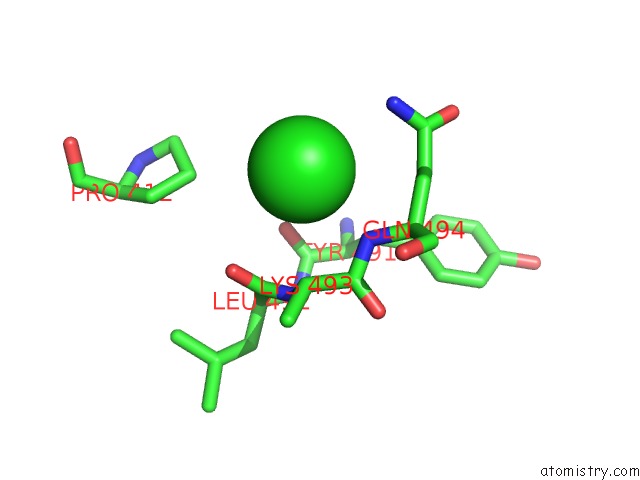
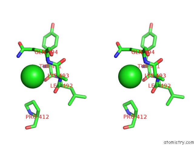
Chlorine binding site 1 out of 4 in 3l5j
Go back to
Chlorine binding site 1 out
of 4 in the Crystal Structure of Fniii Domains of Human GP130 (Domains 4-6)
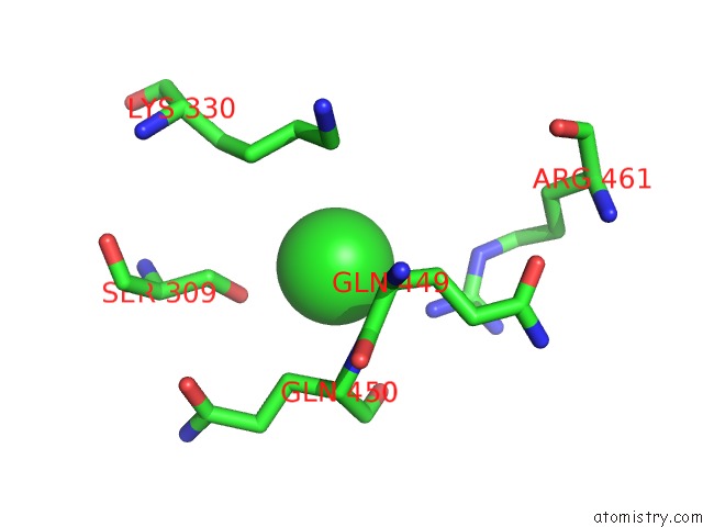
Mono view
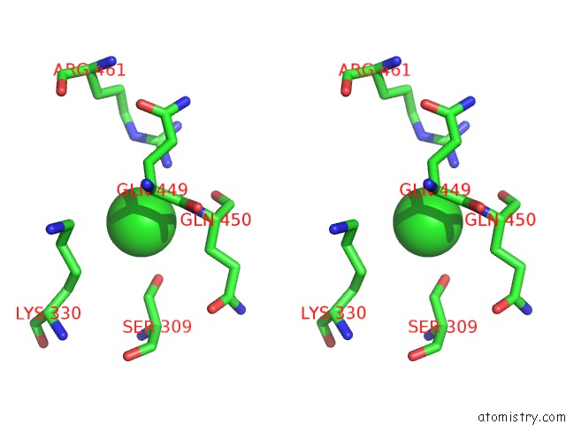
Stereo pair view
Mono view
Stereo pair view
A full contact list of Chlorine with other atoms in the Cl binding
site number 1 of Crystal Structure of Fniii Domains of Human GP130 (Domains 4-6) within 5.0Å range:
|
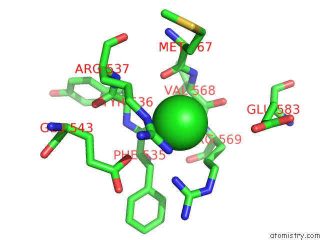
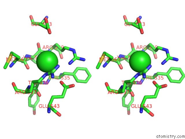
Chlorine binding site 2 out of 4 in 3l5j
Go back to
Chlorine binding site 2 out
of 4 in the Crystal Structure of Fniii Domains of Human GP130 (Domains 4-6)
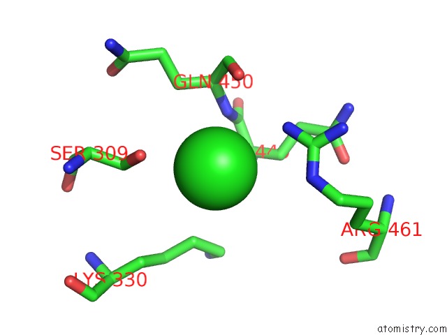
Mono view
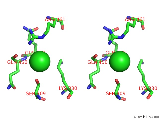
Stereo pair view
Mono view
Stereo pair view
A full contact list of Chlorine with other atoms in the Cl binding
site number 2 of Crystal Structure of Fniii Domains of Human GP130 (Domains 4-6) within 5.0Å range:
|
Chlorine binding site 3 out of 4 in 3l5j
Go back to
Chlorine binding site 3 out
of 4 in the Crystal Structure of Fniii Domains of Human GP130 (Domains 4-6)
Mono view
Stereo pair view
Mono view
Stereo pair view
A full contact list of Chlorine with other atoms in the Cl binding
site number 3 of Crystal Structure of Fniii Domains of Human GP130 (Domains 4-6) within 5.0Å range:
|
Chlorine binding site 4 out of 4 in 3l5j
Go back to
Chlorine binding site 4 out
of 4 in the Crystal Structure of Fniii Domains of Human GP130 (Domains 4-6)
Mono view
Stereo pair view
Mono view
Stereo pair view
A full contact list of Chlorine with other atoms in the Cl binding
site number 4 of Crystal Structure of Fniii Domains of Human GP130 (Domains 4-6) within 5.0Å range:
|
Reference:
Y.Xu,
N.J.Kershaw,
C.S.Luo,
P.Soo,
M.J.Pocock,
P.E.Czabotar,
D.J.Hilton,
N.A.Nicola,
T.P.Garrett,
J.G.Zhang.
Crystal Structure of the Entire Ectodomain of GP130: Insights Into the Molecular Assembly of the Tall Cytokine Receptor Complexes. J.Biol.Chem. V. 285 21214 2010.
ISSN: ISSN 0021-9258
PubMed: 20489211
DOI: 10.1074/JBC.C110.129502
Page generated: Fri Jul 11 07:17:48 2025
ISSN: ISSN 0021-9258
PubMed: 20489211
DOI: 10.1074/JBC.C110.129502
Last articles
Fe in 7VZNFe in 7VYH
Fe in 7VYF
Fe in 7VYP
Fe in 7VYO
Fe in 7VYJ
Fe in 7VYA
Fe in 7VY8
Fe in 7VT2
Fe in 7VXU