Chlorine »
PDB 3l2h-3lcc »
3lbj »
Chlorine in PDB 3lbj: Structure of Human Mdmx Protein in Complex with A Small Molecule Inhibitor
Protein crystallography data
The structure of Structure of Human Mdmx Protein in Complex with A Small Molecule Inhibitor, PDB code: 3lbj
was solved by
G.M.Popowicz,
A.Czarna,
S.Wolf,
T.A.Holak,
with X-Ray Crystallography technique. A brief refinement statistics is given in the table below:
| Resolution Low / High (Å) | 20.00 / 1.50 |
| Space group | I 21 3 |
| Cell size a, b, c (Å), α, β, γ (°) | 80.970, 80.970, 80.970, 90.00, 90.00, 90.00 |
| R / Rfree (%) | 17.4 / 20.6 |
Chlorine Binding Sites:
The binding sites of Chlorine atom in the Structure of Human Mdmx Protein in Complex with A Small Molecule Inhibitor
(pdb code 3lbj). This binding sites where shown within
5.0 Angstroms radius around Chlorine atom.
In total 4 binding sites of Chlorine where determined in the Structure of Human Mdmx Protein in Complex with A Small Molecule Inhibitor, PDB code: 3lbj:
Jump to Chlorine binding site number: 1; 2; 3; 4;
In total 4 binding sites of Chlorine where determined in the Structure of Human Mdmx Protein in Complex with A Small Molecule Inhibitor, PDB code: 3lbj:
Jump to Chlorine binding site number: 1; 2; 3; 4;
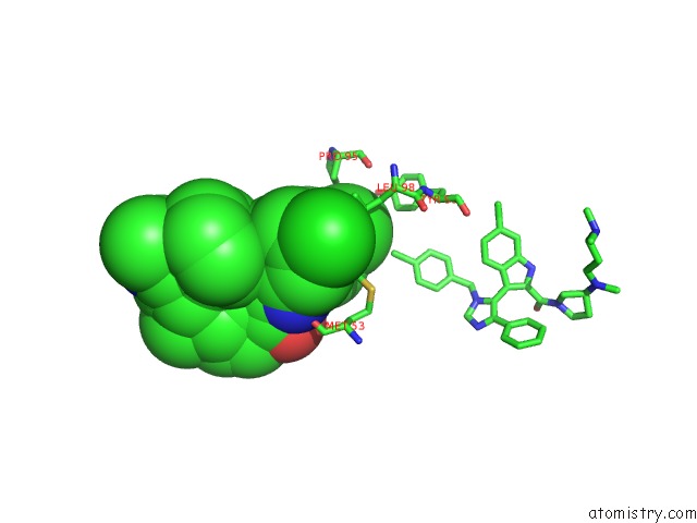
Chlorine binding site 1 out of 4 in 3lbj
Go back to
Chlorine binding site 1 out
of 4 in the Structure of Human Mdmx Protein in Complex with A Small Molecule Inhibitor
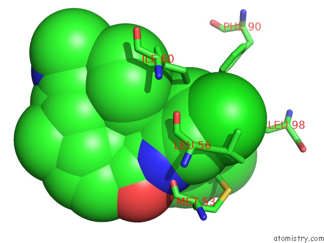
Mono view
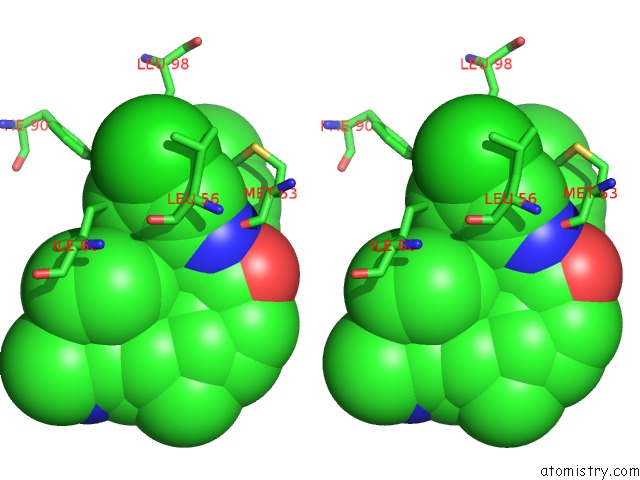
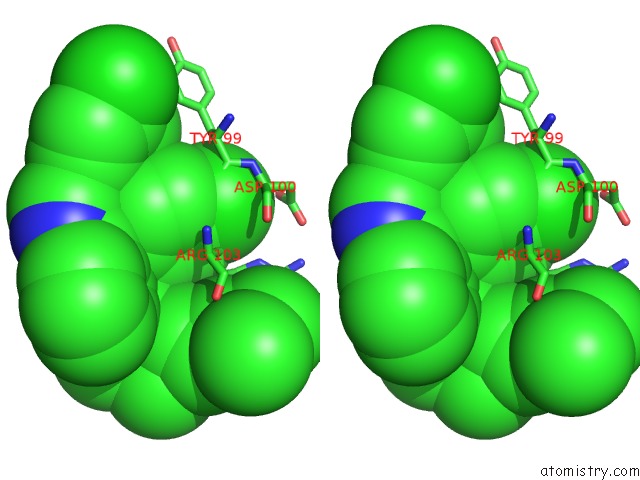
Stereo pair view
Mono view
Stereo pair view
A full contact list of Chlorine with other atoms in the Cl binding
site number 1 of Structure of Human Mdmx Protein in Complex with A Small Molecule Inhibitor within 5.0Å range:
|
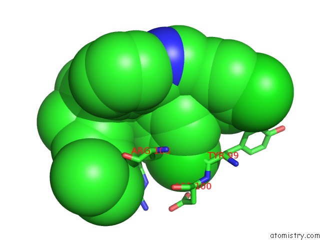
Chlorine binding site 2 out of 4 in 3lbj
Go back to
Chlorine binding site 2 out
of 4 in the Structure of Human Mdmx Protein in Complex with A Small Molecule Inhibitor
Mono view

Stereo pair view
Mono view
Stereo pair view
A full contact list of Chlorine with other atoms in the Cl binding
site number 2 of Structure of Human Mdmx Protein in Complex with A Small Molecule Inhibitor within 5.0Å range:
|
Chlorine binding site 3 out of 4 in 3lbj
Go back to
Chlorine binding site 3 out
of 4 in the Structure of Human Mdmx Protein in Complex with A Small Molecule Inhibitor
Mono view
Stereo pair view
Mono view
Stereo pair view
A full contact list of Chlorine with other atoms in the Cl binding
site number 3 of Structure of Human Mdmx Protein in Complex with A Small Molecule Inhibitor within 5.0Å range:
|
Chlorine binding site 4 out of 4 in 3lbj
Go back to
Chlorine binding site 4 out
of 4 in the Structure of Human Mdmx Protein in Complex with A Small Molecule Inhibitor

Mono view

Stereo pair view
Mono view
Stereo pair view
A full contact list of Chlorine with other atoms in the Cl binding
site number 4 of Structure of Human Mdmx Protein in Complex with A Small Molecule Inhibitor within 5.0Å range:
|
Reference:
G.M.Popowicz,
A.Czarna,
S.Wolf,
K.Wang,
W.Wang,
A.Domling,
T.A.Holak.
Structures of Low Molecular Weight Inhibitors Bound to Mdmx and MDM2 Reveal New Approaches For P53-Mdmx/MDM2 Antagonist Drug Discovery Cell Cycle V. 9 1104 2010.
ISSN: ESSN 1551-4005
PubMed: 20237429
DOI: 10.4161/CC.9.6.10956
Page generated: Fri Jul 11 07:24:47 2025
ISSN: ESSN 1551-4005
PubMed: 20237429
DOI: 10.4161/CC.9.6.10956
Last articles
Fe in 7VYFFe in 7VYP
Fe in 7VYO
Fe in 7VYJ
Fe in 7VYA
Fe in 7VY8
Fe in 7VT2
Fe in 7VXU
Fe in 7VY3
Fe in 7VXP