Chlorine »
PDB 3l2h-3lcc »
3lc2 »
Chlorine in PDB 3lc2: Crystal Structure of Thioacyl-Glyceraldehyde-3-Phosphate Dehydrogenase 1(Gapdh 1) From Methicillin Resistant Staphylococcus Aureus MRSA252
Enzymatic activity of Crystal Structure of Thioacyl-Glyceraldehyde-3-Phosphate Dehydrogenase 1(Gapdh 1) From Methicillin Resistant Staphylococcus Aureus MRSA252
All present enzymatic activity of Crystal Structure of Thioacyl-Glyceraldehyde-3-Phosphate Dehydrogenase 1(Gapdh 1) From Methicillin Resistant Staphylococcus Aureus MRSA252:
1.2.1.12;
1.2.1.12;
Protein crystallography data
The structure of Crystal Structure of Thioacyl-Glyceraldehyde-3-Phosphate Dehydrogenase 1(Gapdh 1) From Methicillin Resistant Staphylococcus Aureus MRSA252, PDB code: 3lc2
was solved by
S.Mukherjee,
D.Dutta,
B.Saha,
A.K.Das,
with X-Ray Crystallography technique. A brief refinement statistics is given in the table below:
| Resolution Low / High (Å) | 20.00 / 2.80 |
| Space group | P 1 21 1 |
| Cell size a, b, c (Å), α, β, γ (°) | 69.057, 103.025, 90.303, 90.00, 109.40, 90.00 |
| R / Rfree (%) | 18.1 / 24.1 |
Chlorine Binding Sites:
The binding sites of Chlorine atom in the Crystal Structure of Thioacyl-Glyceraldehyde-3-Phosphate Dehydrogenase 1(Gapdh 1) From Methicillin Resistant Staphylococcus Aureus MRSA252
(pdb code 3lc2). This binding sites where shown within
5.0 Angstroms radius around Chlorine atom.
In total 3 binding sites of Chlorine where determined in the Crystal Structure of Thioacyl-Glyceraldehyde-3-Phosphate Dehydrogenase 1(Gapdh 1) From Methicillin Resistant Staphylococcus Aureus MRSA252, PDB code: 3lc2:
Jump to Chlorine binding site number: 1; 2; 3;
In total 3 binding sites of Chlorine where determined in the Crystal Structure of Thioacyl-Glyceraldehyde-3-Phosphate Dehydrogenase 1(Gapdh 1) From Methicillin Resistant Staphylococcus Aureus MRSA252, PDB code: 3lc2:
Jump to Chlorine binding site number: 1; 2; 3;
Chlorine binding site 1 out of 3 in 3lc2
Go back to
Chlorine binding site 1 out
of 3 in the Crystal Structure of Thioacyl-Glyceraldehyde-3-Phosphate Dehydrogenase 1(Gapdh 1) From Methicillin Resistant Staphylococcus Aureus MRSA252
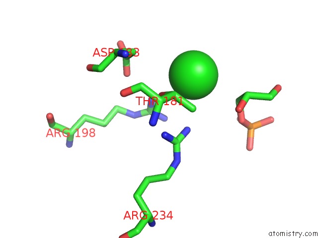
Mono view

Stereo pair view
Mono view
Stereo pair view
A full contact list of Chlorine with other atoms in the Cl binding
site number 1 of Crystal Structure of Thioacyl-Glyceraldehyde-3-Phosphate Dehydrogenase 1(Gapdh 1) From Methicillin Resistant Staphylococcus Aureus MRSA252 within 5.0Å range:
|
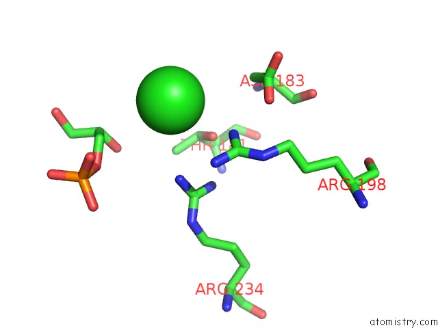
Chlorine binding site 2 out of 3 in 3lc2
Go back to
Chlorine binding site 2 out
of 3 in the Crystal Structure of Thioacyl-Glyceraldehyde-3-Phosphate Dehydrogenase 1(Gapdh 1) From Methicillin Resistant Staphylococcus Aureus MRSA252

Mono view

Stereo pair view
Mono view
Stereo pair view
A full contact list of Chlorine with other atoms in the Cl binding
site number 2 of Crystal Structure of Thioacyl-Glyceraldehyde-3-Phosphate Dehydrogenase 1(Gapdh 1) From Methicillin Resistant Staphylococcus Aureus MRSA252 within 5.0Å range:
|
Chlorine binding site 3 out of 3 in 3lc2
Go back to
Chlorine binding site 3 out
of 3 in the Crystal Structure of Thioacyl-Glyceraldehyde-3-Phosphate Dehydrogenase 1(Gapdh 1) From Methicillin Resistant Staphylococcus Aureus MRSA252
Mono view

Stereo pair view
Mono view
Stereo pair view
A full contact list of Chlorine with other atoms in the Cl binding
site number 3 of Crystal Structure of Thioacyl-Glyceraldehyde-3-Phosphate Dehydrogenase 1(Gapdh 1) From Methicillin Resistant Staphylococcus Aureus MRSA252 within 5.0Å range:
|
Reference:
S.Mukherjee,
D.Dutta,
B.Saha,
A.K.Das.
Crystal Structure of Glyceraldehyde-3-Phosphate Dehydrogenase 1 From Methicillin-Resistant Staphylococcus Aureus MRSA252 Provides Novel Insights Into Substrate Binding and Catalytic Mechanism. J.Mol.Biol. V. 401 949 2010.
ISSN: ISSN 0022-2836
PubMed: 20620151
DOI: 10.1016/J.JMB.2010.07.002
Page generated: Fri Jul 11 07:25:35 2025
ISSN: ISSN 0022-2836
PubMed: 20620151
DOI: 10.1016/J.JMB.2010.07.002
Last articles
Fe in 7VYFFe in 7VYP
Fe in 7VYO
Fe in 7VYJ
Fe in 7VYA
Fe in 7VY8
Fe in 7VT2
Fe in 7VXU
Fe in 7VY3
Fe in 7VXP