Chlorine »
PDB 3lcd-3lnr »
3lk0 »
Chlorine in PDB 3lk0: X-Ray Structure of Bovine SC0067,Ca(2+)-S100B
Protein crystallography data
The structure of X-Ray Structure of Bovine SC0067,Ca(2+)-S100B, PDB code: 3lk0
was solved by
T.H.Charpentier,
D.J.Weber,
P.W.Wilder,
with X-Ray Crystallography technique. A brief refinement statistics is given in the table below:
| Resolution Low / High (Å) | 19.36 / 2.04 |
| Space group | P 1 21 1 |
| Cell size a, b, c (Å), α, β, γ (°) | 28.607, 59.122, 103.998, 90.00, 92.52, 90.00 |
| R / Rfree (%) | 23.5 / 29.1 |
Other elements in 3lk0:
The structure of X-Ray Structure of Bovine SC0067,Ca(2+)-S100B also contains other interesting chemical elements:
| Calcium | (Ca) | 8 atoms |
Chlorine Binding Sites:
The binding sites of Chlorine atom in the X-Ray Structure of Bovine SC0067,Ca(2+)-S100B
(pdb code 3lk0). This binding sites where shown within
5.0 Angstroms radius around Chlorine atom.
In total 2 binding sites of Chlorine where determined in the X-Ray Structure of Bovine SC0067,Ca(2+)-S100B, PDB code: 3lk0:
Jump to Chlorine binding site number: 1; 2;
In total 2 binding sites of Chlorine where determined in the X-Ray Structure of Bovine SC0067,Ca(2+)-S100B, PDB code: 3lk0:
Jump to Chlorine binding site number: 1; 2;
Chlorine binding site 1 out of 2 in 3lk0
Go back to
Chlorine binding site 1 out
of 2 in the X-Ray Structure of Bovine SC0067,Ca(2+)-S100B

Mono view
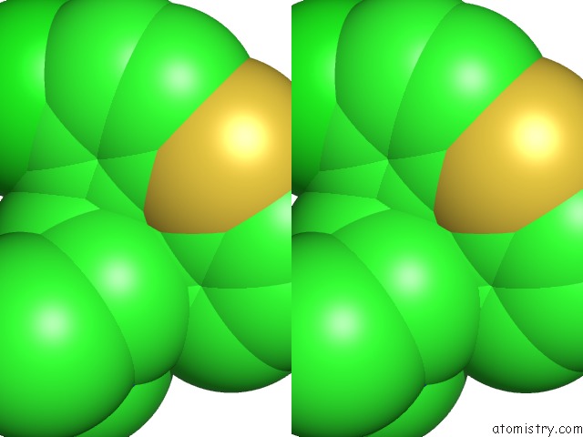
Stereo pair view
Mono view
Stereo pair view
A full contact list of Chlorine with other atoms in the Cl binding
site number 1 of X-Ray Structure of Bovine SC0067,Ca(2+)-S100B within 5.0Å range:
|
Chlorine binding site 2 out of 2 in 3lk0
Go back to
Chlorine binding site 2 out
of 2 in the X-Ray Structure of Bovine SC0067,Ca(2+)-S100B

Mono view

Stereo pair view
Mono view
Stereo pair view
A full contact list of Chlorine with other atoms in the Cl binding
site number 2 of X-Ray Structure of Bovine SC0067,Ca(2+)-S100B within 5.0Å range:
|
Reference:
P.T.Wilder,
T.H.Charpentier,
M.A.Liriano,
K.Gianni,
K.M.Varney,
E.Pozharski,
A.Coop,
E.A.Toth,
A.D.Mackerell,
D.J.Weber.
In Vitro Screening and Structural Characterization of Inhibitors of the S100B-P53 Interaction. Int J High Throughput Screen V.2010 109 2010.
ISSN: ESSN 1179-1381
PubMed: 21132089
DOI: 10.2147/IJHTS.S8210
Page generated: Fri Jul 11 07:28:31 2025
ISSN: ESSN 1179-1381
PubMed: 21132089
DOI: 10.2147/IJHTS.S8210
Last articles
Fe in 2YXOFe in 2YRS
Fe in 2YXC
Fe in 2YNM
Fe in 2YVJ
Fe in 2YP1
Fe in 2YU2
Fe in 2YU1
Fe in 2YQB
Fe in 2YOO