Chlorine »
PDB 3lcd-3lnr »
3lk7 »
Chlorine in PDB 3lk7: The Crystal Structure of Udp-N-Acetylmuramoylalanine-D-Glutamate (Murd) Ligase From Streptococcus Agalactiae to 1.5A
Enzymatic activity of The Crystal Structure of Udp-N-Acetylmuramoylalanine-D-Glutamate (Murd) Ligase From Streptococcus Agalactiae to 1.5A
All present enzymatic activity of The Crystal Structure of Udp-N-Acetylmuramoylalanine-D-Glutamate (Murd) Ligase From Streptococcus Agalactiae to 1.5A:
6.3.2.9;
6.3.2.9;
Protein crystallography data
The structure of The Crystal Structure of Udp-N-Acetylmuramoylalanine-D-Glutamate (Murd) Ligase From Streptococcus Agalactiae to 1.5A, PDB code: 3lk7
was solved by
A.J.Stein,
A.Sather,
G.Shakelford,
A.Joachimiak,
Midwest Center Forstructural Genomics (Mcsg),
with X-Ray Crystallography technique. A brief refinement statistics is given in the table below:
| Resolution Low / High (Å) | 50.00 / 1.50 |
| Space group | C 1 2 1 |
| Cell size a, b, c (Å), α, β, γ (°) | 161.539, 65.045, 52.889, 90.00, 107.52, 90.00 |
| R / Rfree (%) | 18.7 / 20.8 |
Chlorine Binding Sites:
The binding sites of Chlorine atom in the The Crystal Structure of Udp-N-Acetylmuramoylalanine-D-Glutamate (Murd) Ligase From Streptococcus Agalactiae to 1.5A
(pdb code 3lk7). This binding sites where shown within
5.0 Angstroms radius around Chlorine atom.
In total only one binding site of Chlorine was determined in the The Crystal Structure of Udp-N-Acetylmuramoylalanine-D-Glutamate (Murd) Ligase From Streptococcus Agalactiae to 1.5A, PDB code: 3lk7:
In total only one binding site of Chlorine was determined in the The Crystal Structure of Udp-N-Acetylmuramoylalanine-D-Glutamate (Murd) Ligase From Streptococcus Agalactiae to 1.5A, PDB code: 3lk7:
Chlorine binding site 1 out of 1 in 3lk7
Go back to
Chlorine binding site 1 out
of 1 in the The Crystal Structure of Udp-N-Acetylmuramoylalanine-D-Glutamate (Murd) Ligase From Streptococcus Agalactiae to 1.5A
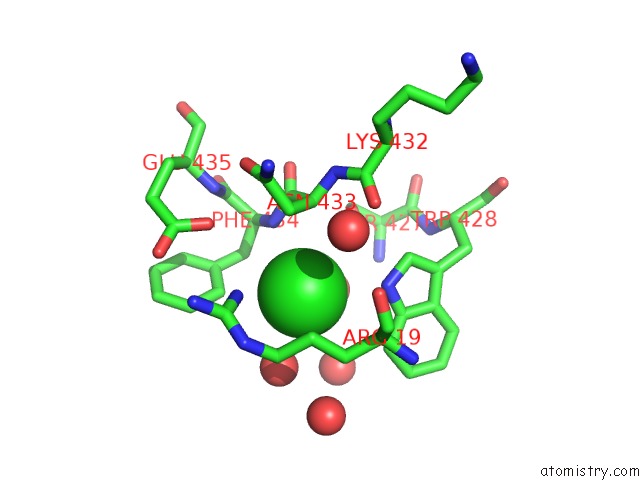
Mono view
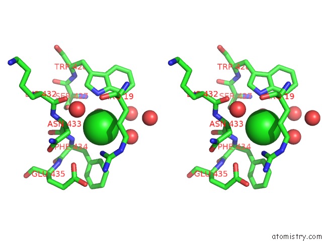
Stereo pair view
Mono view
Stereo pair view
A full contact list of Chlorine with other atoms in the Cl binding
site number 1 of The Crystal Structure of Udp-N-Acetylmuramoylalanine-D-Glutamate (Murd) Ligase From Streptococcus Agalactiae to 1.5A within 5.0Å range:
|
Reference:
A.J.Stein,
A.Sather,
G.Shakelford,
A.Joachimiak.
The Crystal Structure of Udp-N-Acetylmuramoylalanine-D-Glutamate (Murd) Ligase From Streptococcus Agalactiae to 1.5A To Be Published.
Page generated: Fri Jul 11 07:28:40 2025
Last articles
Zn in 4IMXZn in 4IMU
Zn in 4IMS
Zn in 4ILW
Zn in 4IMW
Zn in 4IMT
Zn in 4ILO
Zn in 4ILK
Zn in 4ILX
Zn in 4IL3