Chlorine »
PDB 3lnz-3lz0 »
3lxt »
Chlorine in PDB 3lxt: Crystal Structure of Glutathione S Transferase From Pseudomonas Fluorescens
Protein crystallography data
The structure of Crystal Structure of Glutathione S Transferase From Pseudomonas Fluorescens, PDB code: 3lxt
was solved by
R.Agarwal,
S.K.Burley,
S.Swaminathan,
New York Sgx Researchcenter For Structural Genomics (Nysgxrc),
with X-Ray Crystallography technique. A brief refinement statistics is given in the table below:
| Resolution Low / High (Å) | 44.30 / 1.76 |
| Space group | P 21 21 21 |
| Cell size a, b, c (Å), α, β, γ (°) | 57.681, 57.887, 265.772, 90.00, 90.00, 90.00 |
| R / Rfree (%) | 19.9 / 22.6 |
Chlorine Binding Sites:
The binding sites of Chlorine atom in the Crystal Structure of Glutathione S Transferase From Pseudomonas Fluorescens
(pdb code 3lxt). This binding sites where shown within
5.0 Angstroms radius around Chlorine atom.
In total 7 binding sites of Chlorine where determined in the Crystal Structure of Glutathione S Transferase From Pseudomonas Fluorescens, PDB code: 3lxt:
Jump to Chlorine binding site number: 1; 2; 3; 4; 5; 6; 7;
In total 7 binding sites of Chlorine where determined in the Crystal Structure of Glutathione S Transferase From Pseudomonas Fluorescens, PDB code: 3lxt:
Jump to Chlorine binding site number: 1; 2; 3; 4; 5; 6; 7;
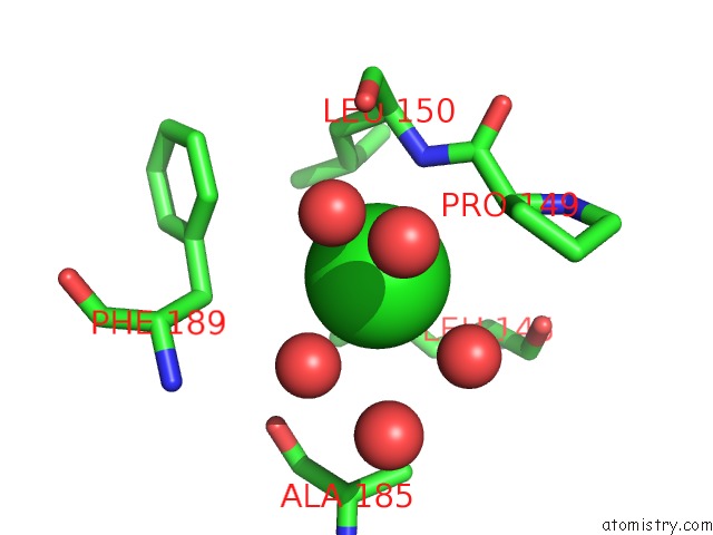
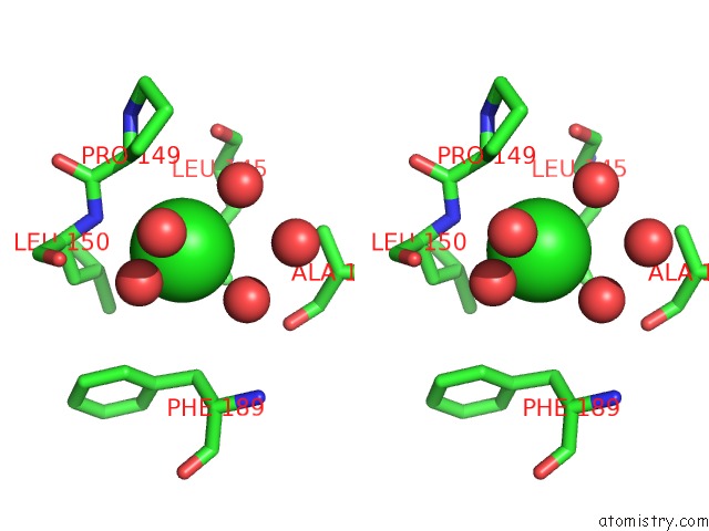
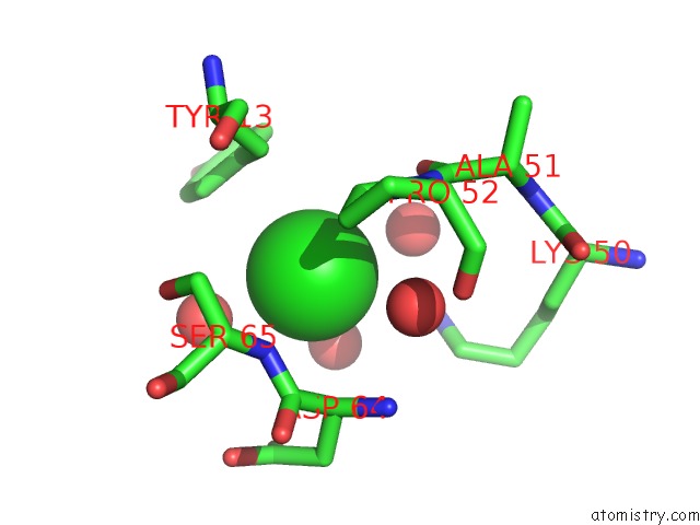
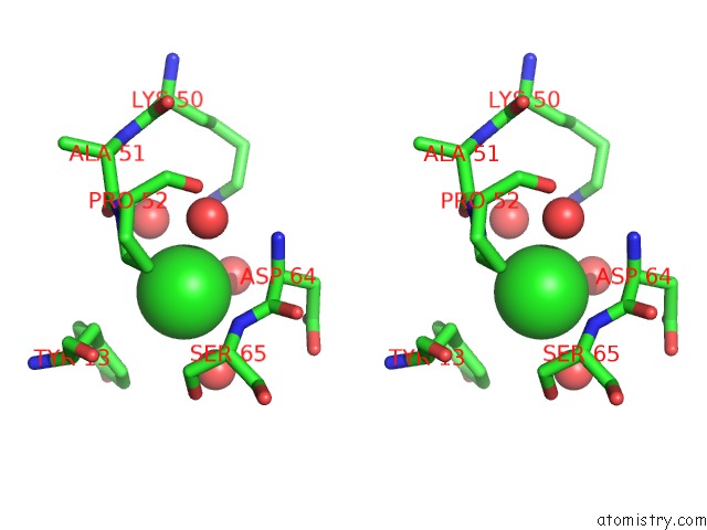
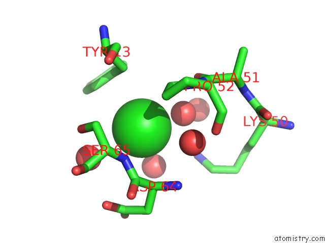
Chlorine binding site 1 out of 7 in 3lxt
Go back to
Chlorine binding site 1 out
of 7 in the Crystal Structure of Glutathione S Transferase From Pseudomonas Fluorescens
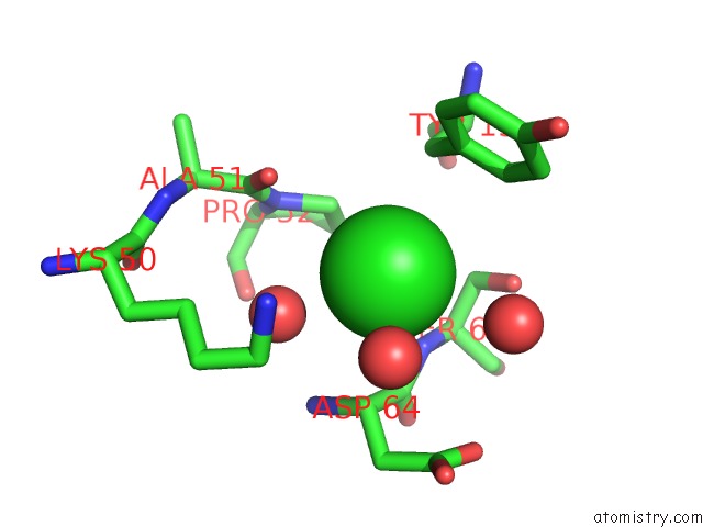
Mono view
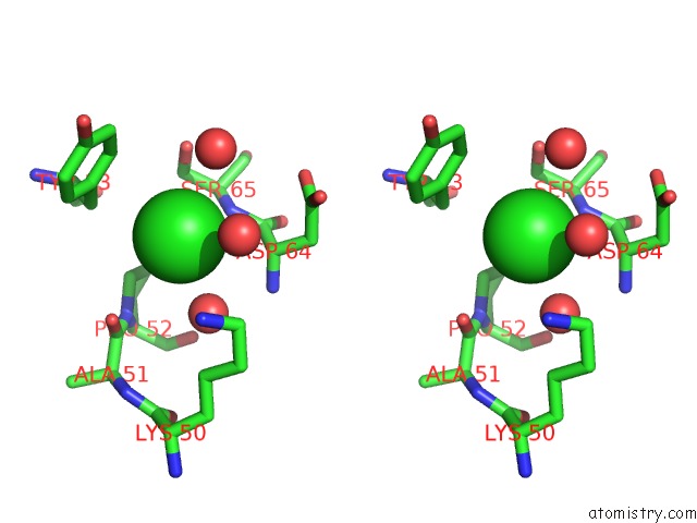
Stereo pair view
Mono view
Stereo pair view
A full contact list of Chlorine with other atoms in the Cl binding
site number 1 of Crystal Structure of Glutathione S Transferase From Pseudomonas Fluorescens within 5.0Å range:
|
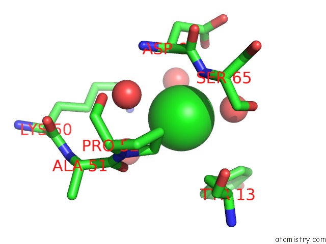
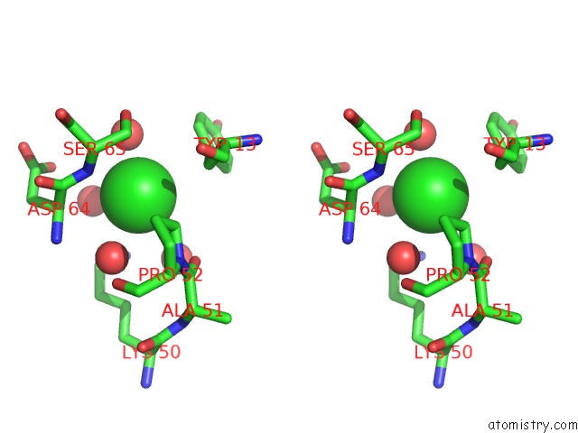
Chlorine binding site 2 out of 7 in 3lxt
Go back to
Chlorine binding site 2 out
of 7 in the Crystal Structure of Glutathione S Transferase From Pseudomonas Fluorescens
Mono view
Stereo pair view
Mono view
Stereo pair view
A full contact list of Chlorine with other atoms in the Cl binding
site number 2 of Crystal Structure of Glutathione S Transferase From Pseudomonas Fluorescens within 5.0Å range:
|
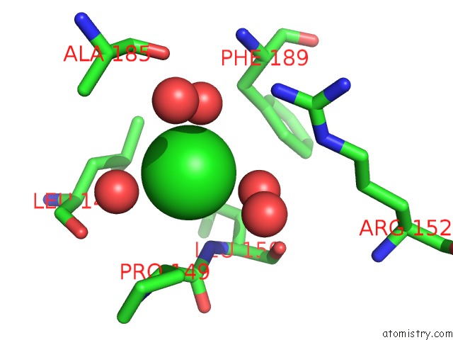
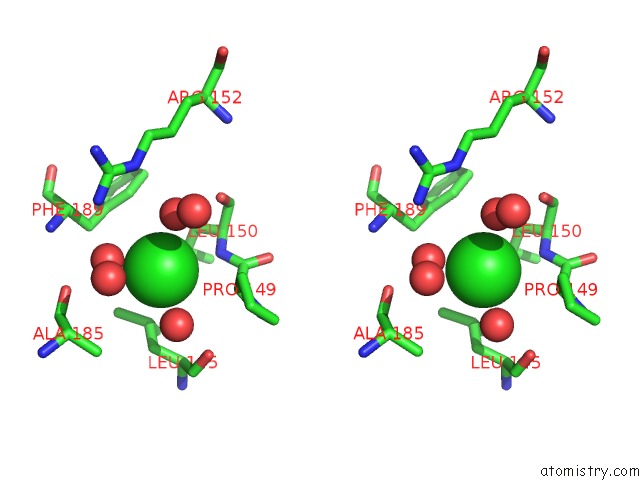
Chlorine binding site 3 out of 7 in 3lxt
Go back to
Chlorine binding site 3 out
of 7 in the Crystal Structure of Glutathione S Transferase From Pseudomonas Fluorescens
Mono view
Stereo pair view
Mono view
Stereo pair view
A full contact list of Chlorine with other atoms in the Cl binding
site number 3 of Crystal Structure of Glutathione S Transferase From Pseudomonas Fluorescens within 5.0Å range:
|
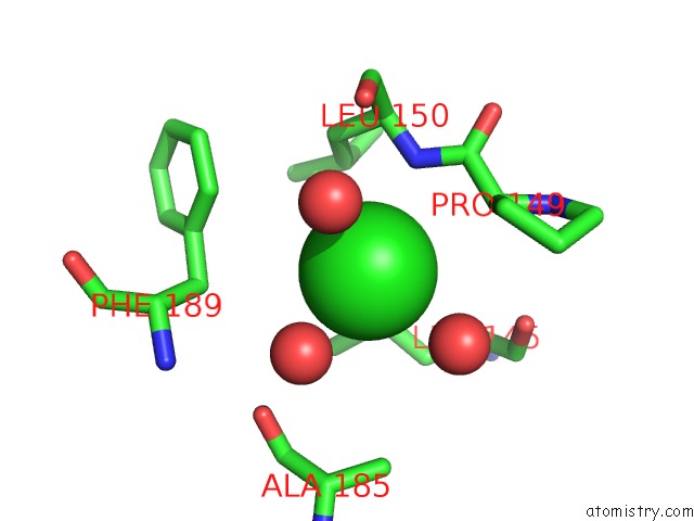
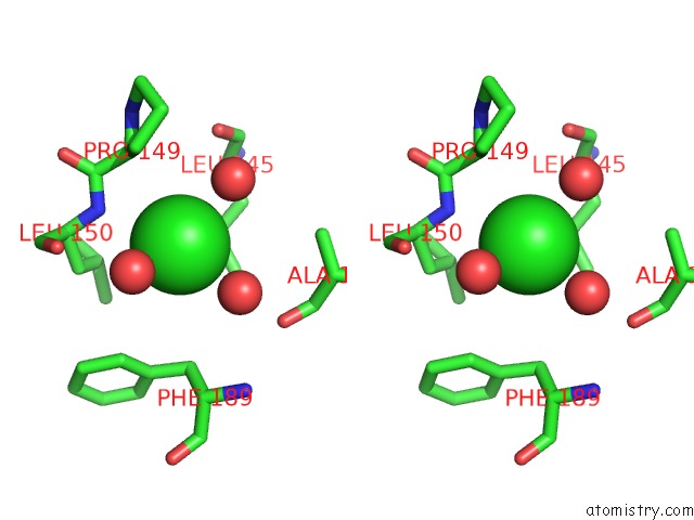
Chlorine binding site 4 out of 7 in 3lxt
Go back to
Chlorine binding site 4 out
of 7 in the Crystal Structure of Glutathione S Transferase From Pseudomonas Fluorescens
Mono view
Stereo pair view
Mono view
Stereo pair view
A full contact list of Chlorine with other atoms in the Cl binding
site number 4 of Crystal Structure of Glutathione S Transferase From Pseudomonas Fluorescens within 5.0Å range:
|
Chlorine binding site 5 out of 7 in 3lxt
Go back to
Chlorine binding site 5 out
of 7 in the Crystal Structure of Glutathione S Transferase From Pseudomonas Fluorescens
Mono view
Stereo pair view
Mono view
Stereo pair view
A full contact list of Chlorine with other atoms in the Cl binding
site number 5 of Crystal Structure of Glutathione S Transferase From Pseudomonas Fluorescens within 5.0Å range:
|
Chlorine binding site 6 out of 7 in 3lxt
Go back to
Chlorine binding site 6 out
of 7 in the Crystal Structure of Glutathione S Transferase From Pseudomonas Fluorescens
Mono view
Stereo pair view
Mono view
Stereo pair view
A full contact list of Chlorine with other atoms in the Cl binding
site number 6 of Crystal Structure of Glutathione S Transferase From Pseudomonas Fluorescens within 5.0Å range:
|
Chlorine binding site 7 out of 7 in 3lxt
Go back to
Chlorine binding site 7 out
of 7 in the Crystal Structure of Glutathione S Transferase From Pseudomonas Fluorescens
Mono view

Stereo pair view
Mono view
Stereo pair view
A full contact list of Chlorine with other atoms in the Cl binding
site number 7 of Crystal Structure of Glutathione S Transferase From Pseudomonas Fluorescens within 5.0Å range:
|
Reference:
R.Agarwal,
S.K.Burley,
S.Swaminathan.
Crystal Structure of Glutathione S Transferase From Pseudomonas Fluorescens To Be Published.
Page generated: Fri Jul 11 07:36:32 2025
Last articles
K in 4EYVK in 4EVY
K in 4EOU
K in 4ETM
K in 4ESK
K in 4ES8
K in 4ERT
K in 4ERD
K in 4ENC
K in 4EK1