Chlorine »
PDB 3lz1-3m98 »
3m13 »
Chlorine in PDB 3m13: Crystal Structure of the LYS265ARG Peg-Crystallized Mutant of Monomeric Sarcosine Oxidase
Enzymatic activity of Crystal Structure of the LYS265ARG Peg-Crystallized Mutant of Monomeric Sarcosine Oxidase
All present enzymatic activity of Crystal Structure of the LYS265ARG Peg-Crystallized Mutant of Monomeric Sarcosine Oxidase:
1.5.3.1;
1.5.3.1;
Protein crystallography data
The structure of Crystal Structure of the LYS265ARG Peg-Crystallized Mutant of Monomeric Sarcosine Oxidase, PDB code: 3m13
was solved by
F.S.Mathews,
Z.-W.Chen,
M.S.Jorns,
with X-Ray Crystallography technique. A brief refinement statistics is given in the table below:
| Resolution Low / High (Å) | 29.20 / 2.10 |
| Space group | P 1 21 1 |
| Cell size a, b, c (Å), α, β, γ (°) | 99.296, 69.298, 111.427, 90.00, 93.40, 90.00 |
| R / Rfree (%) | 18.7 / 24.8 |
Chlorine Binding Sites:
The binding sites of Chlorine atom in the Crystal Structure of the LYS265ARG Peg-Crystallized Mutant of Monomeric Sarcosine Oxidase
(pdb code 3m13). This binding sites where shown within
5.0 Angstroms radius around Chlorine atom.
In total 4 binding sites of Chlorine where determined in the Crystal Structure of the LYS265ARG Peg-Crystallized Mutant of Monomeric Sarcosine Oxidase, PDB code: 3m13:
Jump to Chlorine binding site number: 1; 2; 3; 4;
In total 4 binding sites of Chlorine where determined in the Crystal Structure of the LYS265ARG Peg-Crystallized Mutant of Monomeric Sarcosine Oxidase, PDB code: 3m13:
Jump to Chlorine binding site number: 1; 2; 3; 4;
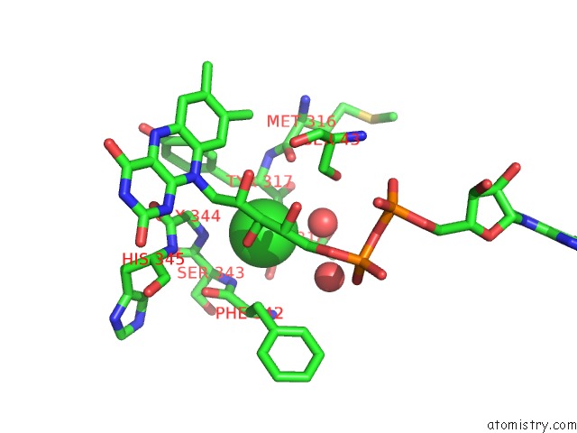
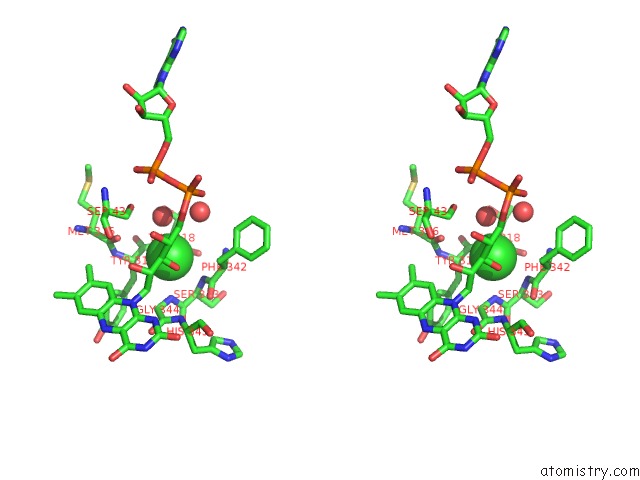
Chlorine binding site 1 out of 4 in 3m13
Go back to
Chlorine binding site 1 out
of 4 in the Crystal Structure of the LYS265ARG Peg-Crystallized Mutant of Monomeric Sarcosine Oxidase
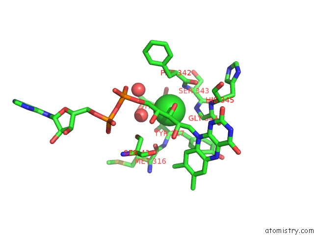
Mono view
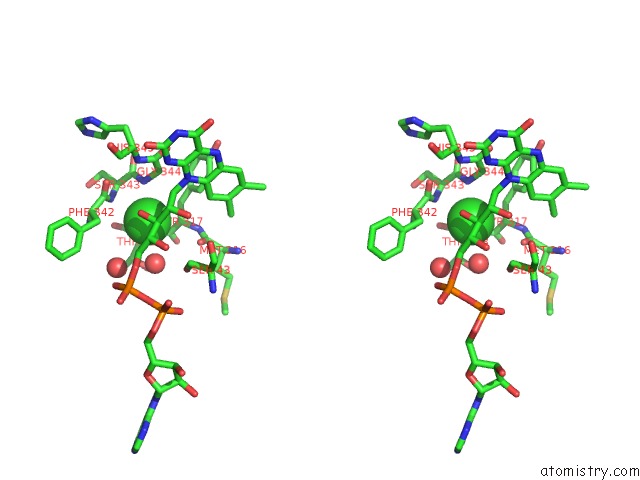
Stereo pair view
Mono view
Stereo pair view
A full contact list of Chlorine with other atoms in the Cl binding
site number 1 of Crystal Structure of the LYS265ARG Peg-Crystallized Mutant of Monomeric Sarcosine Oxidase within 5.0Å range:
|
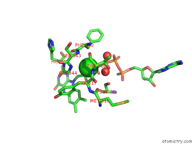
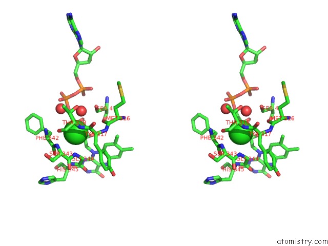
Chlorine binding site 2 out of 4 in 3m13
Go back to
Chlorine binding site 2 out
of 4 in the Crystal Structure of the LYS265ARG Peg-Crystallized Mutant of Monomeric Sarcosine Oxidase
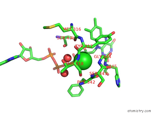
Mono view
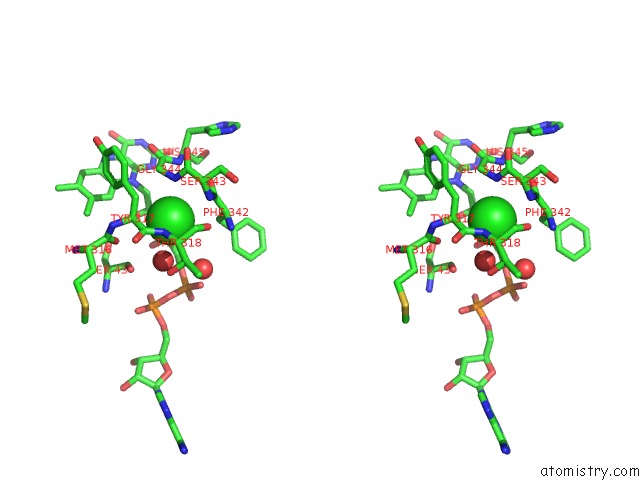
Stereo pair view
Mono view
Stereo pair view
A full contact list of Chlorine with other atoms in the Cl binding
site number 2 of Crystal Structure of the LYS265ARG Peg-Crystallized Mutant of Monomeric Sarcosine Oxidase within 5.0Å range:
|
Chlorine binding site 3 out of 4 in 3m13
Go back to
Chlorine binding site 3 out
of 4 in the Crystal Structure of the LYS265ARG Peg-Crystallized Mutant of Monomeric Sarcosine Oxidase
Mono view
Stereo pair view
Mono view
Stereo pair view
A full contact list of Chlorine with other atoms in the Cl binding
site number 3 of Crystal Structure of the LYS265ARG Peg-Crystallized Mutant of Monomeric Sarcosine Oxidase within 5.0Å range:
|
Chlorine binding site 4 out of 4 in 3m13
Go back to
Chlorine binding site 4 out
of 4 in the Crystal Structure of the LYS265ARG Peg-Crystallized Mutant of Monomeric Sarcosine Oxidase
Mono view
Stereo pair view
Mono view
Stereo pair view
A full contact list of Chlorine with other atoms in the Cl binding
site number 4 of Crystal Structure of the LYS265ARG Peg-Crystallized Mutant of Monomeric Sarcosine Oxidase within 5.0Å range:
|
Reference:
M.S.Jorns,
Z.W.Chen,
F.S.Mathews.
Structural Characterization of Mutations at the Oxygen Activation Site in Monomeric Sarcosine Oxidase. Biochemistry V. 49 3631 2010.
ISSN: ISSN 0006-2960
PubMed: 20353187
DOI: 10.1021/BI100160J
Page generated: Fri Jul 11 07:42:28 2025
ISSN: ISSN 0006-2960
PubMed: 20353187
DOI: 10.1021/BI100160J
Last articles
F in 5TI6F in 5TI4
F in 5TI2
F in 5TDI
F in 5TE8
F in 5TEI
F in 5TD7
F in 5TBP
F in 5TCI
F in 5TBO